
Figure 7. Response to hypercapnic acidosis of an astrocyte from the irRTN.
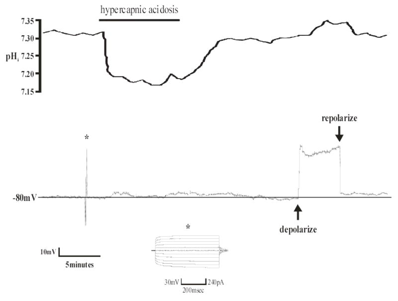
Top panel: pHi trace of an individual astrocyte from the irRTN. Hypercapnic acidosis resulted in a maintained, intracellular acidification that returned towards its initial value upon return to control solution. Bottom panel: Vm trace of the same astrocyte in the top panel. Hypercapnic acidosis (10% CO2, pHo 7.15) caused a depolarization of approximately 5mV, which returned towards normal under control conditions (5% CO2, pHo 7.45). Note that the changes in Vm follow the changes in pHi. Also note the hyperpolarized Vm (− 80mV) and the lack of action potentials, both of which indicate this is an astrocyte. Asterisk indicates a pulse protocol (generated by p-Clamp software) showing that positive current is unable to elicit action potentials, which is further proof that this cell is an astrocyte. At the end of each of these experiments, depolarizing DC current (~ +40mV) was continuously injected via the Dagan amplifier resulting in an intracellular alkalinization (see top panel), which is known as depolarization-induced alkalinization (39). This is additional evidence that this cell is an astrocyte.