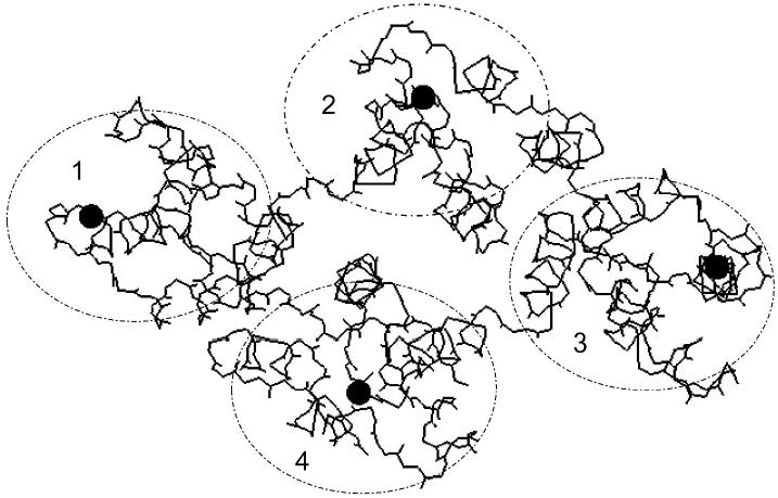
FIGURE 4.

Schematic view of the three-dimensional structure of annexin V-128. The view is downward onto the membrane binding face of the protein. The four domains of the protein are numbered, and their approximate locations are indicated by the ellipses. Black spheres indicate the approximate locations of calcium ions bound to the AB-helix calcium binding sites that have been mutated in this study. The polypeptide backbone is shown as a continuous black line. The location of the N-terminal technetium chelation site can not be seen in this projection because it is on the opposite face of the molecule, beneath the plane of the page. (Structure is based on preliminary coordinates provided by Dr. Barbara Seaton (personal communication.)
