
Figure 6. Inhibition of MMP activity attenuates KCl-induced retinal degeneration.
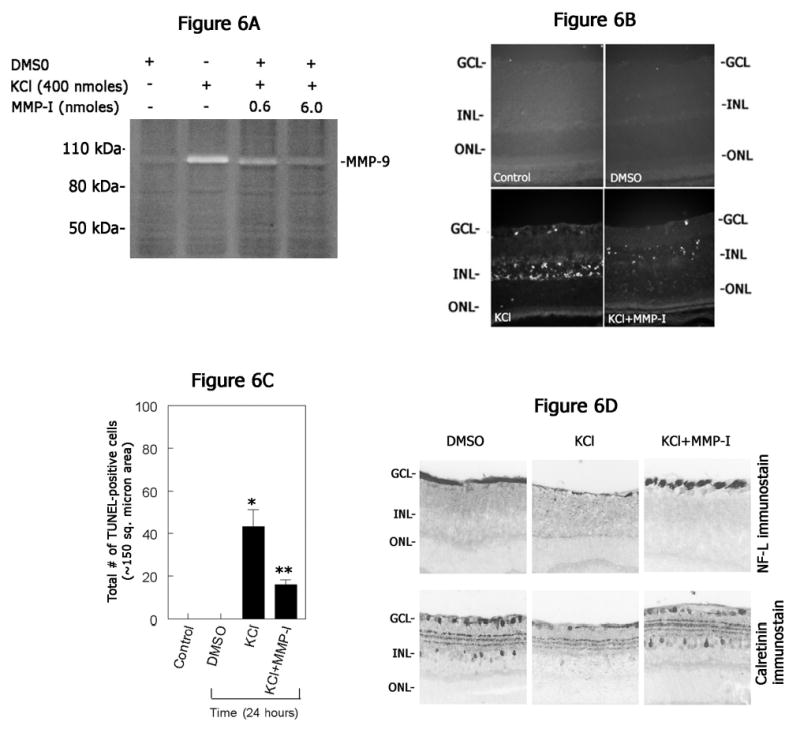
(A) Retinal proteins were extracted at one day after intravitreal injection of KCl (400 nmoles) along with indicated concentrations of MMP inhibitor (MMP-inhibitor-I). Aliquots containing an equal amount of proteins (25 ug) were subjected to gelatin zymography. Note a decrease in KCl-mediated MMP-9 activity after intravitreal injection of MMP inhibitor. (B) Retinal cross sections prepared at one day after intravitreal injection of KCl (400 nmoles) with or without MMP inhibitor (6 nmoles; MMP-inhibitor-I) were subjected to TUNEL assays and compared with un-injected control or vehicle (DMSO)-injected eyes. (C) TUNEL positive cells were counted and plotted as a bar graph. Results indicate a significant reduction in the number of TUNEL positive cells in retinal cross sections after intravitreal injection of KCl along with MMP inhibitor. *P<0.05, controls vs KCl; **P<0.05 KCl vs KCl plus MMP inhibitor; ANOVA, followed by a post hoc Tukey’s test. (D) Retinal cross sections prepared after intravitreal injection of KCl along with or without MMP inhibitor were immunostained with antibodies against neurofilament-light (NF-L) and calretinin. Compared KCl injected eyes, retinal cross sections prepared after intravitreal injection of KCl along with MMP inhibitor showed attenuation in ganglion and amacrine cell loss.