
Fig. 1.
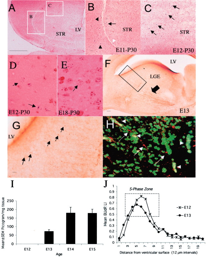
Neurogenesis in the LGE occurs in a dopamine-rich milieu. When E11 mice were exposed to BUdR, labeled cells were found at the lateral margin of the neostriatum (B,arrow) just medial to the external capsule (B, white arrowhead) as well as outside the neostriatal borders on P30 (B, black arrowheads, E11-P30). When E12 mice were exposed to BUdR, labeled cells were found throughout the neostriatum (C, arrows) as well as outside the neostriatum on P30 (E12-P30). The position of labeled cells shown in B and C is indicated bywhite rectangles in A. When a double S-phase labeling method was used, cells labeled with3H-TdR-only were present in the neostriatum on P30 if the S-phase marker injections were administered on E12 (D,arrows, E12-P30) or E18 (E, arrow, E18-P30) but not on E11, confirming that neostriatal neurogenesis began on E12.F, TH-positive axons in the neostriatum (arrow) on E13. The boxed area inF is shown at a higher magnification in Gto illustrate growing tips of TH-positive fibers (G,arrows) within 25–50 μm of the lateral ventricular border. TH-positive axons and growth cones (white arrows) are in close proximity to BUdR-labeled (green) nuclei in the LGE (H). Red blood cells that fluoresce in both the green and red filters appear yellowish orange(H, white arrowheads). Dopamine content of the forebrain was undetectable on E12 and rose dramatically between E12 and E13 (I) coincident with the arrival of TH-positive axons in the LGE (F). BUdR LI decreased between E12 and E13 in the S-phase zone of the LGE (J), coincident with the arrival of dopamine. LV, Lateral ventricle; STR, neostriatum. Scale bars:A, 250 μm; (in B) B,C, 50 μm; (in D) D,E, 5 μm; F, 50 μm; G, 10 μm; H, 20 μm.