
Figure 2. Communication between DRG Axons and OPCs Is Revealed by Time-Lapse Confocal Ca2+ Imaging (Higher Levels of Ca2+ Are Displayed in Warmer Colors).
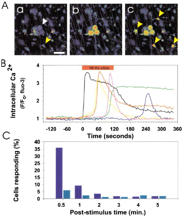
(A) Prior to action potential firing, Ca2+ levels were low in DRG neurons (white arrow) and OPCs (yellow arrow) in coculture (a). Scale bar = 50 μm. Action potentials were induced in DRG axons by electrical stimulation (10 Hz), causing an instantaneous rise in cytoplasmic Ca2+ in the cell body and axons of DRG neurons (b). This was followed by responses in many OPCs (yellow arrows) after several seconds (c). The fluorescence intensity of individual DRG neurons and OPCs in (A) is plotted in (B). 10 Hz electrical stimulation (red bar), cytoplasmic Ca2+ in DRG neurons (black trace), Ca2+ response in OPCs (color traces). No responses to electrical stimulation were seen in OPCs in cultures made without neurons. (C) Incubation with a combination of antagonists of ATP (30 μM suramin) and adenosine receptors (30 μM MRS-1191 and 200 μM DPCPX) inhibited action-potential induced Ca2+ responses in OPCs. A poststimulus time histogram, summarizing the proportion of OPCs responding to action potential firing in the presence (light blue) and absence (dark blue) of these purinergic receptor inhibitors, indicates a significant reduction in short latency (<0.5 min; p < 0.001, χ2, n = 455) and long-latency OPC responses (0.5–5 min; p < 0.02, χ2, n = 455) when purinergic receptors were blocked.