
Figure 3. OPCs at O4− and O4+ Stages Respond to Action Potentials in DRG Axons.
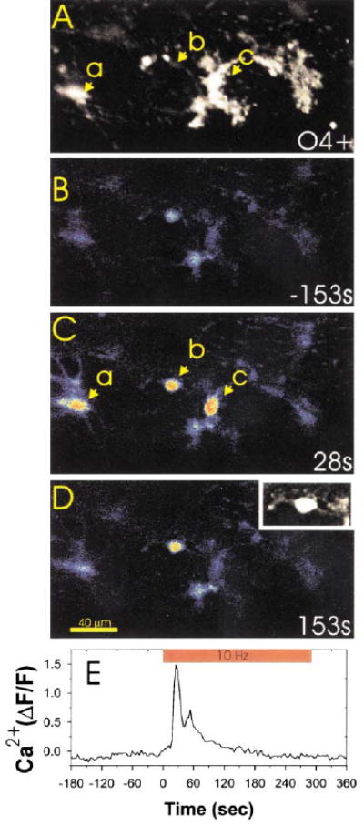
Immunocytochemical staining for the O4 antigen (A) was used after confocal calcium imaging (B–D) to determine the developmental stage at which OPCs responded to action potentials in DRG axons. Three OPCs in this microscope field (a, b, and c) responded to action potentials induced in DRG axons by electrical stimulation. Examination of the same field after O4 staining (A) indicated that OPCs at both the bipolar/O4− stage (cell b and also shown in (D) inset) and O4+ stage (cells a and c in [A] and [C]) responded to axonal firing with large increases in intracellular calcium. Changes in intracellular calcium in cell b are plotted with respect to the time of axonal stimulation in (E). The inset in (D) is an enlargement of cell b filled with the calcium sensitive dye fluo-3, which shows the bipolar cellular morphology more clearly than in the pseudocolor image.