
Fig. 1.
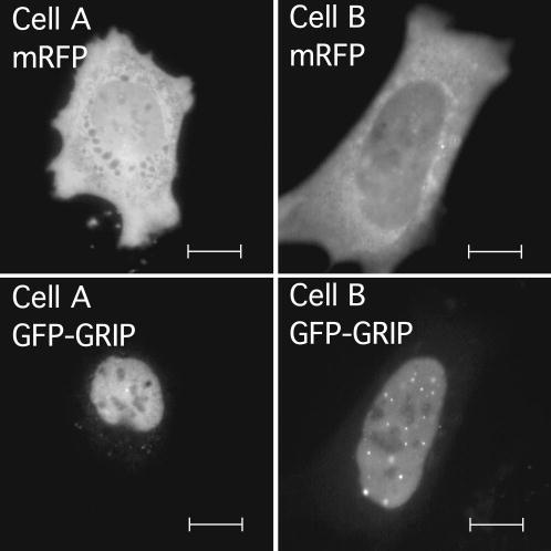
Unbiased cell selection. Pituitary GHFT1-5 cells were cotransfected with vectors encoding mRFP and GFP-GRIP. The living cells expressing the fusion proteins were selected for imaging using the RFP signal. Images of two example cells are shown with mRFP and GFP fluorescence channels displayed separately as labeled. Scale bars are 10 μm in length.