
Figure 2.
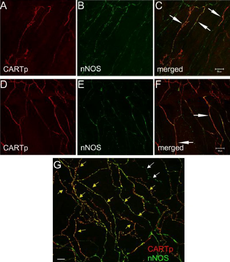
CARTp-IR nerve fibers in suburothelial plexus also express nNOS-immunoreactivity. Confocal fluorescence images of urothelium whole-mounts from adult rats demonstrating CARTp-immunoreactivity (A,D) or nNOS immunoreactivity (B, E) in the same images are shown. Images in C, F and G represent merged images where red staining represents CARTp and green staining represents nNOS. Areas of overlap appear yellow/orange (C, F, white arrows; G, yellow arrows). Some nNOS-IR nerve fibers do not appear to express CARTp-immunoreactivity (G, white arrows) although overlap between CARTp- and nNOS-immunoreactivity is considerable. Calibration bar in C represents 50 μm for A–C. Calibration bar in F represents 50 μm in D–F. Calibration bar represents 100 μm in G.