
Figure 7.
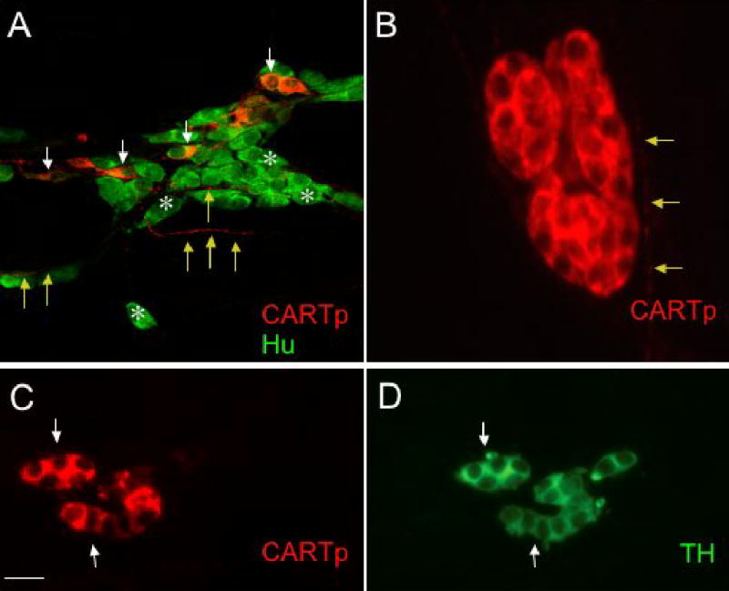
Different populations of CARTp-IR cells are present in detrusor smooth muscle after birth. A. Confocal fluorescence images of cell bodies in detrusor smooth muscle of CARTp+, Hu+ cells (white arrows) as well as numerous neurons (A, Hu+, white asterisks) that lack CARTp-immunoreactivity after birth. CARTp-IR fibers extend among Hu+ cells (A, yellow arrows). In older postnatal and adult rats, CARTp-IR cells that lack a neuronal phenotype are present in detrusor smooth muscle. These CARTp+, Hu- cells (B) are small, rounded cells with large nuclei and a thin rim of cytoplasm. CARTp-IR fibers were observed in close proximity to these cell clusters and (B, yellow arrows). These CARTp+, Hu- cells (C, arrows) express tyrosine hydroxylase (TH) immunoreactivity (D, arrows). Calibration bar represents 30 μm in A, 10 μm in B and 20 μm in C, D.