
Fig. 1.
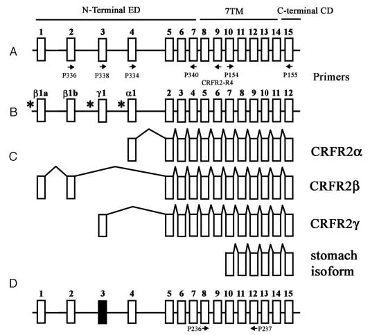
Schematic representation of mammalian CRH-R2 gene structure. A, Human CRH-R2 according to Slominski et al., 2000 (7). B, Human CRH-R2 according to Catalona et al., 2003 (16). C, Alternatively spliced CRH-R2 isoforms α, β, γ (7, 16), and the isoform from stomach (GenBank accession No. E12750; patent no. JP199707289-A). Arrows represent position of PCR primers. Putative promoters sites are shown by * (16). D, Predicted structure of mouse CRH-R2 gene. The structure of mouse gene has been obtained by comparing mouse genomic DNA with published mouse CRH-R2 cDNA and by comparing mouse genomic DNA with human CRHR2 exons (Blast program at http://www.ncbi.nlm.nih.gov/blast). Black box represents mouse exon 3 corresponding to human exon 3 (65% homology). Murine exon 3 contains multiple stop codons and its insertion into the final CRH-R2 mRNA would not produce a functional CRH-R2 receptor. Thus, the equivalent of the human CRH-R2γ isoform will not be expressed in the mouse. Exon 4 has 77% homology at the nucleotide level and 91.3% at the protein level with its human counterpart. Arrows represent position of PCR primers.