
Fig. 3. Expression of MLN64 protein (arrow) in human skin (left) and human and rodent skin cells (right).
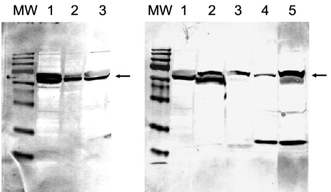
Molecular mass (MW) markers are 180, 130, 73, 54, 48, 35, 24, 16 and 10 kDa. Left: placenta (lanes 1 and 2); skin (lane 3). Right: human SBCE2 (lane 1), WM35 (lane 2), hamster AbC-1 (lane 3) and mouse S-91 (lane 4) melanomas; placenta (lane 5). The amount of protein loaded on gels was 5 and 1 μg for placenta (lanes 1 and 2, respectively) and 20 μg for the skin samples.