
Figure 3.
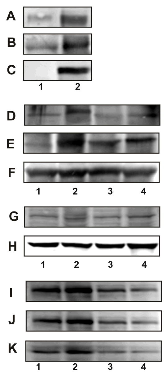
Plasma membrane association of GRP78, NCK, and PAK-2 in 1-LN cells stimulated with α2M*. See EXPERIMENTAL PROCEDURES section for details. Panel A: GRP78 in the GRP78 immunoprecipitate from plasma membrane. Panel B: PAK-2 in the GRP78 immunoprecipitate from plasma membrane. Panel C: NCK in GRP78 immunoprecipitate from plasma membrane. The lanes are: (1) buffer and (2) α2M* (50 pM/10 min). Panel D amd J: Effect of silencing the expression of the GRP78 gene on association of PAK-2. Panel E and K: Effect of silencing the expression of the GRP78 gene on the association of NCK. Panel F: Protein loading control, actin. Panel G and I: Effect of silencing the expression of the GRP78 gene on GRP78 protein levels, and Panel H protein loading control GADPH. The lanes in Panel D, E, F, and G are: (1) buffer; (2) α2M*; (3) dsRNA GRP78 then α2M*; and (4) scrambled dsRNA then α2M*. The lanes in Panel I, J, and K are: (1) buffer; (2) α2M*; (3) dsRNA GRP78 and (4) dsRNA GRP78 then α2M*. The immunoblots shown are representative of three to four independent experiments.