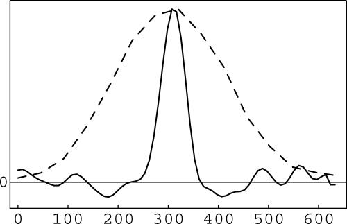
Fig. 7.
Lateral profiles through images of an isolated 50-nm red fluorescent bead, acquired by SSIM (solid trace) and conventional microscopy (dashed trace). (The structure along the baseline in the SSIM profile is caused by noise.) The average FWHM of 100 such SSIM profiles through different beads was 59 nm, a dramatic reduction compared with the 265-nm width for unprocessed conventional microscopy.

