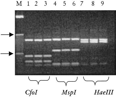
FIG. 3.
Molecular identification of M. mycetomatis from the environment and clinical biopsies. Shown are digests for the specific 26.1A/28.3A PCR products obtained for two clinical isolates (lanes 2 and 3, 5 and 6, and 8 and 9) and a single environmental isolate (lanes 1, 4, and 7). Note that per restriction digest, the patterns are identical. On the left (lane M), the 10-bp molecule sizing ladder is shown, and the 100- and 1,000-bp markers are indicated (arrow).

