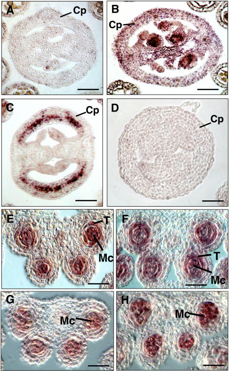
Figure 3.
Expression patterns of TPD1 in the siliques and anthers of wild-type, tpd1, ems1/exs-2, and transgenic plants. A, A transversal section of wild-type silique probed with the TPD1 antisense RNA. B, A transversal section of wild-type silique probed with the EMS1/EXS antisense RNA. C, A transversal section of the transgenic silique probed with the TPD1 antisense RNA. D, A transversal section of wild-type silique probed with the TPD1 sense RNA as a negative control. E, A wild-type anther section probed with the TPD1 antisense RNA. F, A wild-type anther section probed with the EMS1/EXS antisense RNA. G, An ems1 anther section probed with the TPD1 antisense RNA. H, A tpd1 anther section probed with the EMS1/EXS antisense RNA. Cp, Carpel; T, tapetum; Mc, microsporocyte. Bars = 20 μm.

