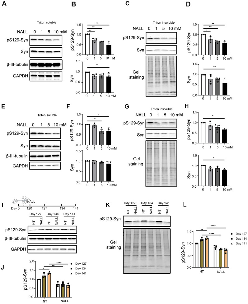
Figure 1. NALL leads to decreased pS129-syn in human dopaminergic neurons with GBA1 mutations.
(A) Western blot showing levels of pS129-syn and total a-Syn (Syn) upon different concentrations of NALL in Triton-soluble faction of GBA1 L444P mutant dopaminergic neurons. β-III-Tubulin and GAPDH were used as loading controls. (B) Quantification of the fold change of decreased pS129-syn (top) and total α-Syn (Syn)(bottom) upon NALL treatment in A. Protein levels normalized toβ-III-Tubulin (n = 3 or 4 independent experiments). (C) Western blot of pS129-syn and total a-Syn (Syn) upon different concentrations of NALL in Triton-insoluble faction of GBA1 L444P mutant dopaminergic neurons. Gel staining was used as a loading control. (D) Quantification of the fold change of decreased pS129-syn (top) and total a-Syn (Syn)(bottom) upon NALL treatment in B. Protein level was normalized with β-III-Tubulin (n = 3 or 4 independent experiments). (E) Western blot of pS129-syn and total a-Syn (Syn) upon different concentrations of NALL treatment in Triton-soluble faction of GBA1 N370S mutant dopaminergic neurons. β-III-Tubulin and GAPDH were used as loading controls. (F) Quantification of the fold change of pS129-syn (top) and total α-Syn (Syn)(bottom) upon NALL treatment in E. The protein level was normalized with β-III-Tubulin (n = 3 independent experiments). (G) Western blot of pS129-syn and total a-Syn (Syn) upon NALL treatment in Triton-insoluble faction of GBA1 N370S mutant dopaminergic neurons. Gel staining was used as a loading control. (H) Quantification of the fold change of pS129-syn (top) and total a-Syn (Syn)(bottom) upon NALL treatment in G. Protein level was normalized with total protein (n = 3 independent experiments). (I) (Top) Schematic illustration of the NALL treatment on dopaminergic neurons. GBA1 L444P mutant neurons were grown for 120 days and treated with or without 10 mM of NALL for another 7, 14, or 21 days. Cells were collected on days 127, 134, or 141. (Bottom) Western blot showing the protein level of pS129-syn with (NALL) or without (NT) NALL treatment in Triton-soluble faction. β-III-Tubulin and GAPDH were used as loading controls. (J) Quantification of the fold change of decreased pS129-syn in I. The protein level was normalized with β-III-Tubulin (n = 3 independent experiments). (K) Western blot of pS129-syn with (NALL) or without (NT) NALL treatment in Triton-insoluble faction of GBA1 L444P mutant dopaminergic neurons. Gel staining was used as a loading control. (L) Quantification of the fold change of pS129-syn in K. Protein level was normalized with total protein (n = 3 independent experiments). All data are represented as mean ± SEM, *p<.05, **p<.01 and ***p<.005 and ****p<.0001.

