



Introduction
In the last few years, Artificial Intelligence (AI) has shot to prominence in the public eye with the recent development of platforms such as ChatGPT. AI is now diffusely incorporated into our everyday human lives without most of us even realising. The expanding use of AI in healthcare shows exciting potential which could transform the future of medicine.
This narrative review is an update on the role of AI in gastrointestinal endoscopy.
What is Artificial Intelligence?
Artificial Intelligence is a broad term that refers to the development of computer systems that can perform tasks that usually would require human intelligence. This is created by programming computers with algorithms that allow them to learn from data, recognise patterns and make decisions without explicit human instruction.1
When it comes to understanding AI systems they can be subdivided into two main categories; Narrow/Weak AI and General/Strong AI. Weak AI are specialised in a particular domain and are only able to perform tasks within their pre- programmed domain.2 For example, the popular household device Alexa is considered a weak AI as it is only able to create responses based on pre-inputted data, meaning it lacks broad understanding and capability to adapt to new non-programmed information. Whereas General/Strong AI is the theorised development of software capable of understanding and utilising knowledge across a broad range of domains.3 Strong AI is envisioned to be able to learn and make decisions independent of trained inputted data, making it scarily comparable to the intrinsic capabilities of the human mind.2,3 The concept of Strong AI is demonstrated in films like Terminator, where Skynet, the military AI gains self-awareness and begins to wage war against humanity. In the current year of 2025, no Strong AI exists.
Generative AI refers to AI that can create new original content such as audio, videos, or text, based on its programmed database, for example Open AI’s ChatGPT.4 Vast amounts of data, over 45 terabytes of text, was inputted into chat GPT’s algorithm enabling it to use this information to learn and create content.5 It is still considered to be a weak form of AI.
AI Terminology
It is important to have a basic understanding of types of AI to help understand and interpret AI models and research studies.
Machine Learning (ML) is a type of AI where the computer uses inputted algorithms to analyse and learn patterns of information from a given set of raw data.1 For example, fitness watches use ML to track and analyse user data, providing insights into physical activity, sleep patterns, and overall health. Similarly our email accounts have the ability to detect incoming junk mail and adequately filter this into a separate inbox (albeit not 100% accurate at all times).
Figure 1: Diagrammatic representation of the definitions of subsets of Artificial Intelligence.1,8.
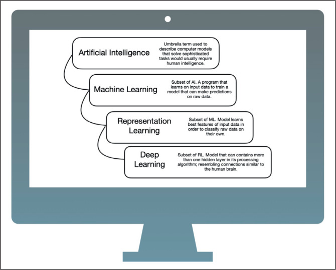
Representation Learning (RL) is a form of ML where the computer uses the best features of inputted data to enable them to classify data on their own. They do this via Artificial Neural Networks (ANNs).6 ANN’s are an AI creation of mathematical models designed to function similarly to the human brains complex connection of signaling neural networks.1
Deep Learning (DL) is a type of RL that uses Deep Neural Networks (DNNs) to form multiple layers of links between vast amounts of data thus enabling it to recognise patterns and automatically make decisions on output.1 For example, facial recognition is a form of RL which enables smart phones to analyse and recognize faces in images or videos. And Spotify, the music streaming service, is an example of DL where the app forms layers and connections to create areas like personalized recommendations, content classification, and music analysis.
In recent years, a main area of interest has leaned heavily on the Large Language Models (LLMs) of AI. These LLMs are types of AI that use Natural Language Processing (NLPs). NLP enables machines to be able to convey information in a human-like response.1,7 Examples of LLMs are Siri, the virtual voice assistant in apple products or ChatGPT that can reply like a human in text form in milliseconds.
AI in the NHS
The UK is considered one of the leading countries in AI research and development. In 2020, the UK ranked third in the world for private investment into AI companies, behind the USA and China. At that time, the government had invested £2.3 billion into AI research across a range of initiatives since 2014.9
In 2019, over £250 million was invested to create the NHS AI Labs for healthcare to help accelerate the safe, ethical and effective development of AI technologies in healthcare.10 The NHS AI Lab aims to collaborate academic research and technology companies to help tackle some of the toughest challenges in NHS healthcare.10 As of March 2023, a total of £123 million had been invested in to 86 different AI technologies used within the NHS.11 The AI in use helps support patients across a range of specialties, and from screening to diagnosis and management of chronic conditions.11 In June 2023, the NHS announced a further £21 million funding for further research into the AI Diagnostic Fund.12 NHS Trusts will be able to bid for funding to accelerate the deployment of the most promising AI tools across hospitals.12
AI in Endoscopy
Within the role of endoscopy; two major AI programs have been studied - Computer assisted detection (CADe) and Computer assisted diagnosis (CADx).13 The role of AI in endoscopy could be hugely beneficial especially considering over 1.5 million endoscopic procedures were performed in 2020 within the NHS alone.14
1. AI in the Oesphagus
Oesophageal cancer is the 7th most common cancer worldwide and the 6th leading cause of cancer-related death.15 Within oesophageal cancer there are two distinct types; oesophageal squamous cell carcinoma (OSCC) being the most common, and oesophageal adenocarcinoma (OAC) following behind.15 Globally the incidence of OAC is rapidly rising in developed, high-income countries, linked to the increasing rate of key risk factors such as obesity, gastro-oesphageal reflux disease (GORD) and Barretts Oesphagus (BO) within this demographic.15 Patients with oesophageal malignancies tend to remain relatively asymptomatic until the tumour has progressed, meaning diagnosis is typically at an advanced stage when survival rate is poor.16 Therefore, the early detection of oesophageal cancer and precancerous lesions is crucial, especially as these early lesions can be treated via endoscopic resection techniques.17
At present, screening for dysplastic BO is composed of targeted biopsies followed by the Seattle protocol in selected individuals.18 This protocol requires physicians to take biopsies every 1-2cm in 4 quadrants and is known to be an inefficient and a time-consuming task.13,18 Endoscopic surveillance then depends on the length of the BO segment and the grade of dysplasia found.19 The critical goal of surveillance programs is for early detection of dysplasia and hence earlier interventions.19,20
AI has used ML models to differentiate between dysplastic and non-neoplastic lesions in numerous studies, with many models showing promising results. Horie et al used over 8,000 endoscopic images to train its AI-CNN network model to be able to detect OSCC and OAC. The model was able to diagnose oesophageal cancer with a sensitivity of 95%, with an impressive 100% detection rate of cancers smaller than 10mm. This model had a remarkably high diagnostic accuracy of 98% in differentiating between superficial and advanced oesophageal cancer.21 Cai et al developed a DNN for CADe using 2,478 endoscopic images with the aim to help improve detection of OSCC. Interestingly, when compared to sixteen endoscopists (senior, mid, and junior level) the system was proven to be superior to both experienced and inexperienced endoscopists in detecting OSCC in endoscopic images, with higher sensitivity and specificity values. The study concluded that their DNN-CAD system could assist in helping endoscopists detect lesions.22
In 2020, Groof et al published one of the first real-time applications of using a CADe system for the detection of neoplasms in patients with Barretts Oesphagus during live gastroscopy in a small pilot study of 20 patients. Of these patients, 10 had nondysplastic BO and 10 patients had confirmed dysplastic BO. The CAD system accurately identified Barrett’s neoplasia at a given level in the Oesophagus with overall 90% accuracy.23
More recently, in 2023 Yuan et al performed a large multi- centre, tandem, double-blind, randomised controlled trial on AI-assisted assessment of early stage OSCC and precancerous lesions. In this study over 5,900 patients were assigned to either AI-first gastroscopy or routine-first gastroscopy. The results showed that AI reduced the miss-rates per lesion from 6.7% to 1.7%.24
At present, AI in the oesophagus shows promise for aiding detection of early cancer or pre-cancerous lesions although it is not available for widespread commercial use yet.
2. AI in the stomach
Worldwide each year approximately one million people are diagnosed with gastric cancer, ranking it 4th for cancer-related mortality globally.25 The prognosis of gastric cancer is largely linked to the stage at which it is diagnosed. Patients with advanced gastric cancer have an extremely poor prognosis, whereas early gastric cancer (EGC) has a 5-year survival rate of more than 90%.26 These differences in figures highlight the importance of early detection and prompt treatment. At present, detection of EGC relies on the direct visualisation of lesions during gastroscopy with the help of some image-enhancing tools.13 EGCs can have variable morphology and hence detection can be challenging even for expert endoscopist. High-quality gastroscopy with full mucosal visualisation is a vital component in improving early detection.
Wu et al created a DNN-AI model using over 9,000 endoscopy images of either gastric cancer or benign lesions to train its model to detect EGC. It then used over 24,000 images for the AI-model to train the model to detect blind spots. The study demonstrated that the trained AI model had a 92.5% accuracy in the detection of EGC in comparison to non-malignancy (with 94.0% sensitivity and 91.0% specificity).27
A large multi-centre study led by Luo et al. Created the GRAIDS model (Gastrointestinal Artificial Intelligence Diagnostic System), for the diagnosis of upper GI cancers using endoscopic images. It used over 1 million images to develop and test the GRAIDS program. The performance of GRAIDS showed a high diagnostic accuracy of 95% with a sensitivity comparable to expert endoscopists. The AI model proved to be superior compared to the endoscopists categorised as competent or trainee.28
In 2020 in Wuhan, China, a randomised control trial with live endoscopy in over 1,800 patients who were undergoing gastroscopy for screening, surveillance, or investigation of symptoms was undertaken. Patients either had an AI- assisted gastroscopy or routine conventional gastroscopy. A conventional gastroscopy showed a neoplasm miss rate of 27.3%, with the AI-assisted endoscopy group being significantly lower with a miss rate of 6.1%. This shows that AI-assisted endoscopy improved the yield of diagnosing gastric neoplasms by endoscopists.29
ENDOANGEL is another example of an AI system showing promising potential for use in gastroscopy. ENDOANGEL, formerly known as WISENSE, was created in 2019 and was aimed at monitoring and reducing rates of blind spots during gastroscopy.30 They then created a DL-AI models, ENDOANGEL-LD (lesion detection), to detect gastric abnormalities and diagnose gastric neoplasms. Over 10,000 patients were enrolled in the study, with results showing in internal and external participants sensitivities of 96.9%/95.6% for detecting gastric lesions and 92.9%/91.7% for diagnosing neoplasms.31
Another key factor when detecting EGC’s is to determine the extent of invasion into the gastric mucosa, which can be challenging in itself. This information is crucial as according to current guidelines, endoscopic resection should only be performed for lesions extending into the mucosal or superficial submucosal layer, regardless of lymph node involvement. For those with deeper invasion the recommended treatment is surgery.32 In 2019, Zhu et al developed a CNN-CAD model using 790 endoscopic images to train the AI system to determine the invasion and depth of gastric cancer. The model was then tested on another 203 images showing an overall accuracy of 89.16%, with higher accuracy and sensitivity when compared with endoscopists, at 71.49% accuracy.33
In summary, AI shows promise in increasing the detection rate of EGCs and to determine the extent of invasion into the gastric mucosa. Cancer Research UK recently awarded a new grant to further develop AI programs for EGC detection showing ongoing dedicated research into endoscopy within the NHS.34 At the current time of writing, no AI models are validated for real-time clinical use. However, at present there is an ongoing multi-centre, randomised, controlled, patient-blinded, trial to test ENDOANGELs new ENDOANGEL-GC (gastric cancer) which will run in real-time endoscopy and combines the functions of blind spot monitoring and lesion detection. The study plans to enroll 30,000 participants from > 20 large-scale primary digestive centres in China.35
3. AI for the detection of H.Pylori
Helicobacter pylori, H.Pylori, is a gram-negative bacterium that specifically colonises the gastric epithelium and is the most common bacterial infection globally, with nearly 50% of the world’s population being infected.36 H.Pylori is well known for its association with gastroduodenal ulcers, gastric carcinoma, or MALT lymphoma.37
At present, testing for H.Pylori during endoscopy requires gastric biopsies with rapid urease test, histology, or culture.38 AI-technology models have been trained to assess and detect the presence of H.Pylori infection during standard endoscopy without the need for a biopsy sample.
In 2018, Itoh et al. created an AI model which used 596 training images for detection of H.Pylori. When the model was tested it showed a sensitivity and specificity of 86.7 % and 86.7 % respectively.39 In 2023, Lin et al designed several CNN networks to test the detection of H.Pylori in images. They concluded their scSE-CatBoost classification CNN-model can achieve a high accuracy for the diagnosis of H. Pylori infection with white light endoscopic image, with an accuracy of 90%, sensitivity 100% and specificity of 81%.40 Whilst AI for assisted detection of H.Pylori is not currently available; studies show that it is entirely possible to detect H.Pylori with visual detection alone in endoscopy. This could in turn reduce the need for unnecessary biopsies and further diagnostic testing, thus widely reducing the cost and carbon footprint.
4. AI in the colon
Every year over 500,000 colonoscopies are performed within the NHS.20 Some for screening purposes, some for diagnostic and others for therapeutic reasons. One of the most common indications for colonoscopy is the investigation of /screening for bowel cancer. Bowel cancer is the 4th most common cancer and the 3nd most common cause of cancer related death worldwide.41 Over 15,000 people die from bowel cancer in UK on an annual basis.42 This figure has been falling since the 1970s, partly due to screening programs and improved treatment techniques.
A key standard in colonoscopy is Post Colonoscopy Colorectal Cancer (PCCRC) rate, that is to say colorectal cancer that develops prior to or seen at the next surveillance colonoscopy.43 It has been shown that an endoscopist’s adenoma detection rate (ADR), the percentage of patients with at least one histologically proven adenoma or carcinoma during colonoscopy, is inversely correlated to PCCRC.43 This highlights that a lower ADR is suggestive of an increased likelihood of missed precancerous adenomas/ malignant lesions. Corley et al meta-analysis of over 300,000 colonoscopies reported that for every 1% increase in ADR the rate of PCCRC drops by 3%.44 Other than ADR, the RCP Joint Advisory Group (JAG) on endoscopy has outlined performance indexes that indicate high quality endoscopy. With regards to colonoscopy these are outlined as >100 procedures by the endoscopist annually, 100% DRE examination, 90% Caecal intubation rate, Adenoma Dectection rate (ADR) 15%, Polyp Retrieval rate 90%, withdrawal time at least 6 minutes, rectal retroversion 90%, adequate bowel preparation in 90%, and targets relating to adverse outcome and patient satisfaction/sedation.43 Of note, European guidelines set a minimal target ADR of 25%.45
To date there have been numerous studies on whether AI leads to an improvement in ADR or not. Rapici et al in Italy showed during a RCT that even in an expert centre ADR was increased from 40.8% in the control to 54.8% with the aid of AI enhanced colonoscopy using Medtronic’s GI Genius.46 Multiple other studies have replicated this data; the caveat however is that they often fail to comment on the level of expertise of the endoscopist. The largest RCT performed to date, by Mangas-Sanjuan et al in Spain, displayed no difference in ADR when their AI program was in the hands of expert endoscopists.47 In this study there was an exceptionally high ADR of 62% within the control group. The wide variance in the performance of endoscopists depending on level of expertise, and various capabilities of the tested AI systems, may be responsible for such discrepancies in reports. And ADRs vary widely, 7 to 53% respectively depending on the endoscopists competence.44 The precursor to ADR is PDR (polyp detection rate) but the two might not be correlated as strongly as one might think. AI increases PDR in nearly all studies one can come across. A substantial proportion of this increase in polyp detection are diminutive polyps (5mm or less).48 They respectively have the lowest pre-test risk of malignant transformation.48 With the practice of excising all detected polyps this leads to an increased, and possibly unnecessary, polyp resection which puts the patient at increased risk of resection related adverse events and increases demand on histopathology services.
Whether this all correlates to an important end-goal outcome, the reduction in PCCRC, that evidence is not available currently. What has been shown is that AI assisted colonoscopy can reduce Adenoma Miss Rate (AMR) on tandem colonoscopies from 37% to 14%.49 The assumption could be made that given a reduction in AMR, there will inevitably be a reduction in PCCRC.
In 2022, M.Ariea et al published their article on a Markov model microsimulation in the Lancet. They hypothesised the effect of AI assisted colonoscopy in a screening program compared to non-augmented colonoscopy within the US population. Their model theorised that on a national level AI assisted colonoscopy would lead to 7,194 less cases of CRC and 2,089 less deaths secondary to CRC and a yearly cost saving of $290 million dollars.50 However many of the authors in this paper had conflicts of interests as they were consultants to companies developing AI programs for endoscopy.
Then there is the second possible capability of AI - CADx. AI point of care diagnosis. The ability to competently differentiate between precancerous adenomas and other types of polyp such as hyperplastic or sessile at the time of visualisation. Current standards stated by PIVI (The American Society for Gastrointestinal Endoscopy Preservation and Incorporation of Valuable Endoscopic Innovation) recommend that if a greater than 90% negative predicative value at visual diagnosis for adenoma, then a diagnose and leave strategy could be implemented in the sigmoid colon, and a resect and discard strategy could be employed in the remaining colon for diminutive polyps (<5mm).51 The ability to implement a dissect and discard approach based of effective CADx is of perceived great economic benefit as a number of diminutive polyps may not require pathological analysis. Multiple studies have shown that AI-augmented colonoscopy surpasses PIVI standards and supports a dissect and discard/diagnose and leave strategy.52,53 There is suggestion there is reluctance to employ these strategies by endoscopists as they inherently come with perceived increased medical-legal risk.54 It has however been shown to improve confidence of endoscopists attempting to visually diagnose polyps at resection.52 Expert endoscopists trained in optical diagnosis can exceed these PIVI standards.55 By implementing CADe technology this could help expert endoscopists feel confident enough in their own ability, enabling them to adopt a resect and discard/diagnose and leave approach when AI is in concordance with their decision, i.e. a second pair of expert eyes in agreeance. Currently these strategies are scarcely used worldwide. AI may be the tool that overcomes the barriers to entry with this approach, increasing endoscopist performance and confidence.
In June 2023, the UK government awarded a fund of 2.5million for use of Medtronic’s GI genius AI endoscopy system. This is called the NAIAD trial (Nationwide study of Artificial Intelligence in Adenoma Detection for colonoscopy). It is led by Kings College London, and taking place over a 2-year period, it will involve 4,000 colonoscopy patients in 20 different hospitals across England. So far, this is the largest study on use of AI in gastroenterology within the NHS.56
At present, there are commercially available AI systems that can be used as an adjunct during colonoscopy to increase adenoma detection. GI Genius of Medtronic (Ireland), EndoBrain of Cybernet (Japan) and Endo-Aid of Olympus (Japan) as a few examples.
Other than detecting and diagnosing polyps with the assistance of AI there are other areas undergoing evaluation. AI programs have been developed that can assist endoscopists in determining quality of colonoscopy by providing immediate feedback on certain aspects. These include percentage of bowel mucosa visualised, grading of bowel preparation, quality of image resolution, the appropriateness of bowel distension and the adequacy of the withdrawal speed.57 With regards to analysing the degree of mucosal invasion AI has been shown to distinguish non-invasive and superficially invasive neoplasms from invasive neoplasms with an accuracy of 91%.58
Investigation into IBD has also been promising with AI programs being shown to be able to detect persistent histological inflammation based on endoscopic images with an accuracy of 91%.59
5. AI in Small Capsule Endoscopy (SCE)
Capsule Endoscopy is a less invasive procedure compared to gastroscopy and colonoscopy. The patient swallows a vitamin sized capsule that contains a camera, light array and transmitter which is connected to an external storage device worn by the patient. The capsule then travels the length of the digestive tract capturing images along the way, most importantly of the small intestine. The small bowel can be visualised in entirety during successful double balloon enteroscopy but SCE has much fewer inherent risks, is more tolerable by patients and achieves complete imaging of the small bowel in a greater percentage of studies.60 Its major drawback being the inability at present to implement treatment at time of visualisation. The indications for SCE are wide. In the small bowel SCEs are the first line investigation for suspected small bowel bleeds, have use in the detection of small bowel tumors and aiding the diagnosis and evaluation of Crohn’s and Coeliac disease.61 They also have their benefits in the oesophagus and large bowel, particularly when sedation for slightly more invasive procedures would be deemed risky or delays procedure due to logistical reasons.61
The duration from ingestion to complete passage through the small bowel varies greatly during SCE. The average time taken to reach and pass through the ileocecal valve can be expected to be around 4-6 hours.62 This results in the acquisition of approximately 50,000 images, all of which require detailed review. Quality standards recommend expert readers set more than 45 minutes aside to review one single investigation, with regular breaks to avoid fatigue and associated lapses in concentration.63
Reducing the time required for clinician analysis and improvement in diagnostic accuracy are the two perceivable outcomes from well working AI in SCE. By reducing the number of redundant/duplicate images the clinician is required to review and presenting images determined to contain pathology a clinician’s time can be effectively prioritised.
Most studies analyse the ability of an AI program to detect a particular type of lesion. Aoiki et al showed an accuracy of 90.8% for the detection of erosions and ulcers, while Fan et al. found an accuracy of 95.2%.64,65 Yuan et al displayed an accuracy of 98% for AI assisted detection of polyps.66 As AI programs become even more sophisticated it is hoped that accuracy will continue to improve. Ideally the AI programs in question would be proficient in detecting multiple abnormalities. For this there are fewer studies to draw from but Ding et al showed a sensitivity of 99.9% for AI augmented reading vs 74.6% for gastroenterologists alone (field of 20) in over 5000 investigations with a mean reading time of 5.9 minutes vs 96.6 minutes.67 This study failed to comment on the gastroenterologists expertise level and their sensitivity of 74.6% is remarkably low compared to expert standards. It does however highlight the benefits AI can achieve in readers with lower expertise.
The introduction of various capsule systems with inbuilt artificial intelligence reached the market. The Navicam SB of ANX Robotica (USA), OMOM HD of Jinshan (China) and the Mirocam of IntroMedic (S.Korea) are some commercially available systems.
Limitations for AI
The growing research in AI in endoscopy is exciting for future practice, however it also comes with new challenges and limitations that need to be recognised. The major limitation inherent to AI, is hallucinations. This is a computer term relevant to AI but it can be understood as errors on the programs part. For example, a chatbot or computer tool can produce impressive and seemingly correct answers however these are non-sensical and not based on any training data. In healthcare this may translate as an incorrectly diagnosed malignant lesion based on no recognised input algorithm. In endoscopy, this could lead to unnecessary biopsies or intervention, or conversely result in ignoring pathology of concern. Thankfully, there are ways to reduce AI hallucinations. The major factors that impact the occurrence of hallucinations are discussed here.
Modern AI systems develop CNNs from a catalogue of input data that teaches it to the desired output. We live in the age of big data where AI systems have the potential to be exposed to an enormous amount of input training data. However, that is not always for the better. There is evidence to suggest that past a certain point, the more data imputed into the AI for training, the more likely that the algorithm will be specific to the training set and not applicable to a wider population.68 Overtraining of the program leads to overfitting. This can result in an increased chance of incorrect responses to new data. I.e. the quality of the training data is just as important as the volume of data used for training. Training an AI program with diverse, high quality raw data is one approach to reducing the rate of hallucinations.
Another way incorrect responses arise is from inappropriate use. Clinicians will be required to ensure that they are using any AI technology in the setting that it was validated for use in. For example, a program developed with the ability to detect sites of bleeding within the large bowel during colonoscopy. It would be inappropriate for a clinician to use this software to aid detection of bleeding during gastroscopy unless validated to do so. While the software may still recognise and detect bleeding sites during gastroscopy, if it has not been validated to do so, then the risk of missed or inappropriately labelled pathology is unknown. It may be that AI programs will be intelligent enough to know when they are being used inappropriately and therefore direct clinicians to this nature.
Generally, DLL models output a response without explaining the decision process and diagnosis basis. With any given response interpreters are unable to ascertain how AI has arrived at that particular response – this issue is referred to as the ‘Blackbox’ of AI. With this, physicians are directed to place their trust in AI programs, while these programs are unable to explain how or why they arrived at a particular outcome. Clinicians are unable to fact check the logic behind the program’s response. If an adverse outcome were to manifest from an incorrect response that a clinician acts on – it raises the issue as to who is responsible, clinician or computer? In our opinion, the clinician has the ultimate responsibility for clinical care and decisions made. They should be aware of the potential shortcomings of their investigative tools. Its for this reason, AI programs will always require a degree of clinician oversight. AI programs should assist clinicians but be overruled when the clinician’s clinical acumen would lead them to take a differing response. A way to help overcome the ‘Blackbox’ issue is through developing AI systems in which the program can logistically explain how it arrived at a specific response. Within endoscopy, ENDOANGEL-ED (Explainable Diagnosis) has been developed for EGC.30 It presents its responses identifying six key analysis areas that aided its decision making. AI programs with inbuilt functions of this nature will help to improve trust and confidence in produced responses.
One perceived benefit of AI in endoscopy is the view that when computerised CADx and CADe systems are in place this will improve standardisation across the field, as it makes endoscopy a less operator dependent procedure. This can also help compensate for human errors that occur due to many factors such as fatigue, stress, or inattention. Whilst this is an agreeable point, there is the antagonistic view of - are we then making endoscopists reliant and dependent on AI? Will this limit their skill set and reduce their role in autonomous decisions in patient care or will it simply increase the speed of their learning curve?
Summary
AI-assisted endoscopy has the potential to improve both patient outcomes and simultaneously reduce clinician workload. While the clinician will never be replaced, these systems will endeavor to act as an assistant with capabilities that a human cannot possess, such as heightened visual acuity, advanced pattern recognition and increased processing speed. Clinicians will be required to educate themselves on new innovative technologies and use them effectively for their intended purpose, whilst also acknowledging the limitations and challenges created when working with AI.
Footnotes
UMJ is an open access publication of the Ulster Medical Society (http://www.ums.ac.uk).
REFERENCES
- 1.Google for Developers [Internet] Machine Learning Glossary; [[cited 2024 May 27]]. Available from: https://developers.google.com/machine-learning/glossary. [Google Scholar]
- 2.Strong AI vs. Weak AI: What’s the Difference? Built In [Internet] 2025. [[cited 2024 May 26]]. Available from: https://builtin.com/artificial-intelligence/strong-ai-weak-ai.
- 3.What Is Strong AI? | IBM [Internet] 2021. [[cited 2024 May 26]]. Available from: https://www.ibm.com/topics/strong-ai.
- 4.Informa. What is Generative AI? Everything You Need to Know. Enterprise AI [Internet] 2024. [[cited 2024 May 26]]. Available from: https://www.techtarget.com/searchenterpriseai/definition/generative-AI.
- 5.McKinsey & Company. What is ChatGPT, DALL-E, and generative AI? [Internet] 2024. [[cited 2024 May 26]]. Available from: https://www.mckinsey.com/featured-insights/mckinsey-explainers/what-is-generative-ai.
- 6.Pannala R, Krishnan K, Melson J, Parsi MA, Schulman AR, Sullivan S, et al. Artificial intelligence in gastrointestinal endoscopy. VideoGIE. 2020;5(12):598–613. doi: 10.1016/j.vgie.2020.08.013. [DOI] [PMC free article] [PubMed] [Google Scholar]
- 7.Thirunavukarasu AJ, Ting DSJ, Elangovan K, Gutierrez L, Tan TF, Ting DSW. Large language models in medicine. Nat Med. 2023;29(8):1930–40. doi: 10.1038/s41591-023-02448-8. [DOI] [PubMed] [Google Scholar]
- 8.Goodfellow I, Bengio Y, Courville A. Deep learning. Cambridge, MA: MIT Press; 2016. [Google Scholar]
- 9.GOV.UK [Internet] National AI Strategy - HTML version. [[cited 2024 May 28]]. Available from: https://www.gov.uk/government/publications/national-ai-strategy/national-ai-strategy-html-version.
- 10.GOV.UK [Internet] News Story. Health Secretary announces £250 million investment in artificial intelligence. [[cited 2024 May 26]]. Available from: https://www.gov.uk/government/news/health-secretary-announces-250-million-investment-in-artificial-intelligence.
- 11.GOV.UK [Internet] Press Release. Thousands of patients to benefit from quicker diagnosis and more accurate tests from ground-breaking AI research. 2023. [[cited 2024 May 26]]. Available from: https://www.gov.uk/government/news/thousands-of-patients-to-benefit-from-quicker-diagnosis-more-accurate-tests-from-ground-breaking-ai-research.
- 12.GOV.UK [Internet] Press Release. £21 million to roll out artificial intelligence across the NHS. 2023. [[cited 2024 May 26]]. Available from: https://www.gov.uk/government/news/21-million-to-roll-out-artificial-intelligence-across-the-nhs.
- 13.Hajjar AE, Rey JF. Artificial intelligence in gastrointestinal endoscopy: general overview. Chin Med J. 2020;133(3):326–34. doi: 10.1097/CM9.0000000000000623. [DOI] [PMC free article] [PubMed] [Google Scholar]
- 14.Ravindran S, Thomas-Gibson S, Bano M, Robinson E, Jenkins A, Marshall S, et al. National census of UK endoscopy services 2021. Frontline Gastroenterol. 2022;13(6):463–70. doi: 10.1136/flgastro-2022-102157. [DOI] [PMC free article] [PubMed] [Google Scholar]
- 15.Liu C, Ma Y, Qin Q, Wang P, Luo Y, Xu P, et al. Epidemiology of esophageal cancer in 2020 and projections to 2030 and 2040. Thorac Cancer. 2022;14(1):3–11. doi: 10.1111/1759-7714.14745. [DOI] [PMC free article] [PubMed] [Google Scholar]
- 16.Short MW, Burgers KG, Fry VT. Esophageal cancer. Am Family Physician. 2017;95(1):22–8. [PubMed] [Google Scholar]
- 17.Pimentel-Nunes P, Libânio D, Bastiaansen BAJ, Bhandari P, Bisschops R, Bourke MJ, et al. Endoscopic submucosal dissection for superficial gastrointestinal lesions: European Society of Gastrointestinal Endoscopy (ESGE) Guideline – Update 2022. Endoscopy. 2022;54(6):591–622. doi: 10.1055/a-1811-7025. [DOI] [PubMed] [Google Scholar]
- 18.Shaheen NJ, Falk GW, Iyer PG, Souza RF, Yadlapati RH, Sauer BG, et al. Diagnosis and management of Barrett’s esophagus: An updated ACG Guideline. Am J Gastroenterol |[ACG] 2022;117(4):559–87. doi: 10.14309/ajg.0000000000001680. [DOI] [PMC free article] [PubMed] [Google Scholar]
- 19.Wang KK, Sampliner RE, Practice Parameters Committee of the American College of Gastroenterology Updated guidelines 2008 for the diagnosis, surveillance and therapy of Barrett’s esophagus. Am J Gastroenterol |[ACG] 2008;103(3):788–97. doi: 10.1111/j.1572-0241.2008.01835.x. [DOI] [PubMed] [Google Scholar]
- 20.Peleg N, Ollech JE, Shamah S, Sapoznikov B. Seattle Protocol Is More Effective in Detection of Dysplasia Compared to Technology-Assisted Targeted Biopsies in Patients with Barrett’s esophagus. J Clin Med. 2023 Mar 28;12(7):2544. doi: 10.3390/jcm12072544. [DOI] [PMC free article] [PubMed] [Google Scholar]
- 21.Horie Y, Yoshio T, Aoyama K, Yoshimizu S, Horiuchi Y, Ishiyama A, et al. Diagnostic outcomes of esophageal cancer by artificial intelligence using convolutional neural networks. Gastrointestinal Endosc. 2019;89(1):25–32. doi: 10.1016/j.gie.2018.07.037. [DOI] [PubMed] [Google Scholar]
- 22.Cai SL, Li B, Tan WM, Niu XJ, Yu HH, Yao LQ, et al. Using a deep learning system in endoscopy for screening of early esophageal squamous cell carcinoma (with video) Gastrointestinal Endosc. 2019;90(5):745–753.e2. doi: 10.1016/j.gie.2019.06.044. [DOI] [PubMed] [Google Scholar]
- 23.De Groof AJ, Struyvenberg MR, Fockens KN, van der Putten J, van der Sommen F, Boers TG, et al. Deep learning algorithm detection of Barrett’s neoplasia with high accuracy during live endoscopic procedures: a pilot study (with video) Gastrointestinal Endosc. 2020;91(6):1242–50. doi: 10.1016/j.gie.2019.12.048. [DOI] [PubMed] [Google Scholar]
- 24.Yuan XL, Liu W, Lin YX, Deng QY, Gao YP, Wan L, et al. Effect of an artificial intelligence-assisted system on endoscopic diagnosis of superficial oesophageal squamous cell carcinoma and precancerous lesions: a multicentre, tandem, double-blind, randomised controlled trial. Lancet Gastroenterol Hepatol. 2024;9(1):34–44. doi: 10.1016/S2468-1253(23)00276-5. [DOI] [PubMed] [Google Scholar]
- 25.Ilic M, Ilic I. Epidemiology of stomach cancer. World J Gastroenterol. 2022;28(12):1187–203. doi: 10.3748/wjg.v28.i12.1187. [DOI] [PMC free article] [PubMed] [Google Scholar]
- 26.Song Z, Wu Y, Yang J, Yang D, Fang X. Progress in the treatment of advanced gastric cancer. Tumour Biol. 2017 Jul 1;39(7):1010428317714626. doi: 10.1177/1010428317714626. [DOI] [PubMed] [Google Scholar]
- 27.Wu L, Zhou W, Wan X, Zhang J, Shen L, Hu S, et al. A deep neural network improves endoscopic detection of early gastric cancer without blind spots. Endoscopy. 2019;51(6):522–31. doi: 10.1055/a-0855-3532. [DOI] [PubMed] [Google Scholar]
- 28.Luo H, Xu G, Li C, He L, Luo L, Wang Z, et al. Real-time artificial intelligence for detection of upper gastrointestinal cancer by endoscopy: a multicentre, case-control, diagnostic study. Lancet Oncology. 2019;20(12):1645–54. doi: 10.1016/S1470-2045(19)30637-0. [DOI] [PubMed] [Google Scholar]
- 29.Wu L, Shang R, Sharma P, Zhou W, Liu J, Yao L, et al. Effect of a deep learning-based system on the miss rate of gastric neoplasms during upper gastrointestinal endoscopy: a single-centre, tandem, randomised controlled trial. Lancet Gastroenterol Hepatol. 2021;6(9):700–8. doi: 10.1016/S2468-1253(21)00216-8. [DOI] [PubMed] [Google Scholar]
- 30.Dong Z, Wang J, Li Y, Deng Y, Zhou W, Zeng X, et al. Explainable artificial intelligence incorporated with domain knowledge diagnosing early gastric neoplasms under white light endoscopy. NPJ Digit Med. 2023;6(1):1–9. doi: 10.1038/s41746-023-00813-y. [DOI] [PMC free article] [PubMed] [Google Scholar]
- 31.Wu L, Xu M, Jiang X, He X, Zhang H, Ai Y, et al. Real-time artificial intelligence for detecting focal lesions and diagnosing neoplasms of the stomach by white-light endoscopy (with videos) Gastrointest Endosc. 2022;95(2):269–80.e6. doi: 10.1016/j.gie.2021.09.017. [DOI] [PubMed] [Google Scholar]
- 32.Banks M, Graham D, Jansen M, Gotoda T, Coda S, Pietro M di, et al. British Society of Gastroenterology guidelines on the diagnosis and management of patients at risk of gastric adenocarcinoma. Gut. 2019;68(9):1545–75. doi: 10.1136/gutjnl-2018-318126. [DOI] [PMC free article] [PubMed] [Google Scholar]
- 33.Zhu Y, Wang QC, Xu MD, Zhang Z, Cheng J, Zhong YS, et al. Application of convolutional neural network in the diagnosis of the invasion depth of gastric cancer based on conventional endoscopy. Gastrointest Endosc. 2019;89(4):806–15.e1. doi: 10.1016/j.gie.2018.11.011. [DOI] [PubMed] [Google Scholar]
- 34.Cambridge University Hospitals NHS Foundation Trust, di Pietro M, [Internet] Using AI to detect stomach cancer. 2024. [[cited 2024 May 26]]. Available from: https://www.cuh.nhs.uk/news/using-ai-to-detect-stomach-cancer/
- 35.Dong Z, Zhu Y, Du H, Wang J, Zeng X, Tao X, et al. The effectiveness of a computer-aided system in improving the detection rate of gastric neoplasm and early gastric cancer: study protocol for a multi-centre, randomized controlled trial. Trials. 2023;24(1):323. doi: 10.1186/s13063-023-07346-5. [DOI] [PMC free article] [PubMed] [Google Scholar]
- 36.Hooi JK, Lai WY, Ng WK, Suen MM, Underwood FE, Tanyingoh D, et al. Global prevalence of Helicobacter Pylori infection: systematic review and meta-analysis. Gastroenterol. 2017;153(2):420–9. doi: 10.1053/j.gastro.2017.04.022. [DOI] [PubMed] [Google Scholar]
- 37.Usui Y, Taniyama Y, Endo M, Koyanagi YN, Kasugai Y, Oze I, et al. Helicobacter pylori, Homologous-Recombination Genes, and Gastric Cancer. N Engl J Med. 2023;388(13):1181–90. doi: 10.1056/NEJMoa2211807. [DOI] [PubMed] [Google Scholar]
- 38.Polk DB, Peek RM. Helicobacter pylori: gastric cancer and beyond. Nat Rev Cancer. 2010;10(6):403–14. doi: 10.1038/nrc2857. [DOI] [PMC free article] [PubMed] [Google Scholar]
- 39.Itoh T, Kawahira H, Nakashima H, Yata N. Deep learning analyzes Helicobacter pylori infection by upper gastrointestinal endoscopy images. Endosc Int Open. 2018;06(2):E139–44. doi: 10.1055/s-0043-120830. [DOI] [PMC free article] [PubMed] [Google Scholar]
- 40.Lin CH, Hsu PI, Tseng CD, Chao PJ, Wu IT, Ghose S, et al. Application of artificial intelligence in endoscopic image analysis for the diagnosis of a gastric cancer pathogen-Helicobacter pylori infection. Sci Rep. 2023;13(1):13380. doi: 10.1038/s41598-023-40179-5. [DOI] [PMC free article] [PubMed] [Google Scholar]
- 41.Rawla P, Sunkara T, Barsouk A. Epidemiology of colorectal cancer: incidence, mortality, survival, and risk factors. Prz Gastroenterol. 2019;14(2):89–103. doi: 10.5114/pg.2018.81072. [DOI] [PMC free article] [PubMed] [Google Scholar]
- 42.Ng OH, Lunevicius R, Arthur JD. Rising incidence of colorectal cancer in individuals younger than 50 years and increasing mortality from rectosigmoid cancer in England. Colorectal Dis. 2021;23(10):2637–46. doi: 10.1111/codi.15819. [DOI] [PubMed] [Google Scholar]
- 43.Rees CJ, Gibson ST, Rutter MD, Baragwanath P, Pullan R, Feeney M, et al. UK key performance indicators and quality assurance standards for colonoscopy. Gut. 2016;65(12):1923–9. doi: 10.1136/gutjnl-2016-312044. [DOI] [PMC free article] [PubMed] [Google Scholar]
- 44.Corley DA, Jensen CD, Marks AR, Zhao WK, Lee JK, Doubeni CA, et al. Adenoma detection rate and risk of colorectal cancer and death. N Engl J Med. 2014;370(14):1298–306. doi: 10.1056/NEJMoa1309086. [DOI] [PMC free article] [PubMed] [Google Scholar]
- 45.Kaminski M, Thomas-Gibson S, Bugajski M, Bretthauer M, Rees C, Dekker E, et al. Performance measures for lower gastrointestinal endoscopy: a European Society of Gastrointestinal Endoscopy (ESGE) Quality Improvement Initiative. Endoscopy. 2017;49(04):378–97. doi: 10.1055/s-0043-103411. [DOI] [PubMed] [Google Scholar]
- 46.Repici A, Badalamenti M, Maselli R, Correale L, Radaelli F, Rondonotti E, et al. Efficacy of real-time computer-aided detection of colorectal neoplasia in a randomized trial. Gastroenterol. 2020;159(2):512–520.e7. doi: 10.1053/j.gastro.2020.04.062. [DOI] [PubMed] [Google Scholar]
- 47.Mangas-Sanjuan C, de-Castro L, Cubiella J, Díez-Redondo P, Suárez A, Pellisé M, et al. Role of artificial intelligence in colonoscopy detection of advanced neoplasias : A randomized trial. Ann Intern Med. 2023;176(9):1145–52. doi: 10.7326/M22-2619. [DOI] [PubMed] [Google Scholar]
- 48.Duvvuri A, Chandrasekar VT, Srinivasan S, Narimiti A, Dasari C, Nutalapati V, et al. Risk of colorectal cancer and cancer related mortality after detection of low-risk or high-risk adenomas, compared with no adenoma, at index colonoscopy: A systematic review and meta-analysis. Gastroenterol. 2021;160(6):1986–1996.e3. doi: 10.1053/j.gastro.2021.01.214. [DOI] [PubMed] [Google Scholar]
- 49.Kamba S, Tamai N, Saitoh I, Matsui H, Horiuchi H, Kobayashi M, et al. Reducing adenoma miss rate of colonoscopy assisted by artificial intelligence: a multicenter randomized controlled trial. J Gastroenterol. 2021;56(8):746–57. doi: 10.1007/s00535-021-01808-w. [DOI] [PubMed] [Google Scholar]
- 50.Areia M, Mori Y, Correale L, Repici A, Bretthauer M, Sharma P, et al. Cost-effectiveness of artificial intelligence for screening colonoscopy: a modelling study. Lancet Digital Health. 2022;4(6):e436–44. doi: 10.1016/S2589-7500(22)00042-5. [DOI] [PubMed] [Google Scholar]
- 51.Rex DK, Kahi C, O’Brien M, Levin TR, Pohl H, Rastogi A, et al. The American Society for Gastrointestinal Endoscopy PIVI (Preservation and Incorporation of Valuable Endoscopic Innovations) on real-time endoscopic assessment of the histology of diminutive colorectal polyps. Gastrointest Endosc. 2011;73(3):419–22. doi: 10.1016/j.gie.2011.01.023. [DOI] [PubMed] [Google Scholar]
- 52.Hassan C, Balsamo G, Lorenzetti R, Zullo A, Antonelli G. Artificial intelligence allows leaving-in-situ colorectal polyps. Clin Gastroenterol Hepatol. 2022;20(11):2505–13.e4. doi: 10.1016/j.cgh.2022.04.045. [DOI] [PubMed] [Google Scholar]
- 53.Rondonotti E, Hassan C, Tamanini G, Antonelli G, Andrisani G, Leonetti G, et al. Artificial intelligence-assisted optical diagnosis for the resect- and-discard strategy in clinical practice: the Artificial intelligence BLI Characterization (ABC) study. Endoscopy. 2023;55(1):14–22. doi: 10.1055/a-1852-0330. [DOI] [PubMed] [Google Scholar]
- 54.Rex DK. It’s not lack of evidence holding back resect and discard. Gastrointest Endosc. 2022;96(6):1033–5. doi: 10.1016/j.gie.2022.08.002. [DOI] [PubMed] [Google Scholar]
- 55.Abu Dayyeh BK, Thosani N, Konda V, Wallace MB, Rex DK, Chauhan SS, et al. ASGE Technology Committee systematic review and meta-analysis assessing the ASGE PIVI thresholds for adopting real-time endoscopic assessment of the histology of diminutive colorectal polyps. Gastrointest Endosc. 2015;81(3):502.e1–502.e16. doi: 10.1016/j.gie.2014.12.022. [DOI] [PubMed] [Google Scholar]
- 56.King’s College Hospital NHS Trust. Nationwide study of artificial intelligence in adenoma detection for colonoscopy. Report No.: NCT05870332. [Internet]. Clinical Trials.gov. 2024. [[cited 2024 Jan 1]]. Available from: https://clinicaltrials.gov/study/NCT05870332.
- 57.Thakkar S, Carleton NM, Rao B, Syed A. Use of artificial intelligence-based analytics from live colonoscopies to optimize the quality of the colonoscopy examination in real time: proof of concept. Gastroenterol. 2020;158(5):1219–21.e2. doi: 10.1053/j.gastro.2019.12.035. [DOI] [PMC free article] [PubMed] [Google Scholar]
- 58.Luo X, Wang J, Han Z, Yu Y, Chen Z, Huang F, et al. Artificial intelligence–enhanced white-light colonoscopy with attention guidance predicts colorectal cancer invasion depth. Gastrointest Endosc. 2021;94(3):627–38.e1. doi: 10.1016/j.gie.2021.03.936. [DOI] [PubMed] [Google Scholar]
- 59.Maeda Y, Kudo SE, Mori Y, Misawa M, Ogata N, Sasanuma S, et al. Fully automated diagnostic system with artificial intelligence using endocytoscopy to identify the presence of histologic inflammation associated with ulcerative colitis (with video) Gastrointest Endosc. 2019;89(2):408–15. doi: 10.1016/j.gie.2018.09.024. [DOI] [PubMed] [Google Scholar]
- 60.Chen X, Ran ZH, Tong JL. A meta-analysis of the yield of capsule endoscopy compared to double-balloon enteroscopy in patients with small bowel diseases. World J Gastroenterol. 2007;13(32):4372–8. doi: 10.3748/wjg.v13.i32.4372. [DOI] [PMC free article] [PubMed] [Google Scholar]
- 61.Pennazio M, Rondonotti E, Despott EJ, Dray X, Keuchel M, Moreels T, et al. Small-bowel capsule endoscopy and device-assisted enteroscopy for diagnosis and treatment of small-bowel disorders: European Society of Gastrointestinal Endoscopy (ESGE) Guideline – Update 2022. Endoscopy. 2023;55(1):58–95. doi: 10.1055/a-1973-3796. [DOI] [PubMed] [Google Scholar]
- 62.Flemming J, Cameron S. Small bowel capsule endoscopy: Indications, results, and clinical benefit in a University environment. Medicine. 2018;97(14):e0148. doi: 10.1097/MD.0000000000010148. [DOI] [PMC free article] [PubMed] [Google Scholar]
- 63.Rondonotti E, Pennazio M, Toth E, Koulaouzidis A. How to read small bowel capsule endoscopy: a practical guide for everyday use. Endosc Int Open. 2020;8(10):E1220–4. doi: 10.1055/a-1210-4830. [DOI] [PMC free article] [PubMed] [Google Scholar]
- 64.Aoki T, Yamada A, Aoyama K, Saito H, Tsuboi A, Nakada A, et al. Automatic detection of erosions and ulcerations in wireless capsule endoscopy images based on a deep convolutional neural network. Gastrointest Endosc. 2019;89(2):357–63.e2. doi: 10.1016/j.gie.2018.10.027. [DOI] [PubMed] [Google Scholar]
- 65.Fan S, Xu L, Fan Y, Wei K, Li L. Computer-aided detection of small intestinal ulcer and erosion in wireless capsule endoscopy images. Phys Med Biol. 2018;63(16):165001. doi: 10.1088/1361-6560/aad51c. [DOI] [PubMed] [Google Scholar]
- 66.Yuan Y, Meng MQ. Deep learning for polyp recognition in wireless capsule endoscopy images. Med Phys. 2017;44(4):1379–89. doi: 10.1002/mp.12147. [DOI] [PubMed] [Google Scholar]
- 67.Ding Z, Shi H, Zhang H, Zhang H, Tian S, Zhang K, et al. Artificial intelligence-based diagnosis of abnormalities in small-bowel capsule endoscopy. Endoscopy. 2023;55(1):44–51. doi: 10.1055/a-1881-4209. [DOI] [PubMed] [Google Scholar]
- 68.Ying X. An overview of overfitting and its solutions. J Phys: Conf Ser. 2019;1168:022022. doi: 10.1088/1742-6596/1168/2/022022. [DOI] [Google Scholar]