



Are we ready for Human Neuroscience?
Modern neuroscience began with Paul Broca’s discovery of language cortex based on observations that lesions in the human brain disrupt normal cognitive functions. Subsequently, Wilder Penfield went on to map the functional areas of the human cerebral cortex through intraoperative stimulation to localize language, memory, and movements. Subsequent mechanistic studies to understand brain function beyond localization have primarily been advanced over the last few decades in animal models. While revealing fundamental principles about the formation and function of the neural circuit, this has left a gap in our understanding of the regulators in higher functional domains such as language and higher sensory processing. Uncovering their genetic, cellular, and molecular basis of cognitive specializations observed in humans (Box 1) has prompted renewed interest in studying the human brain. However, studies of the human brain are challenging due to the lack of scalable tools to manipulate cell behavior or gene expression, and confounding variables of individuality.
What makes us human?
Important insights into brain function have been gained from animal models. Mice offer the opportunity to genetically access and manipulate neural circuits. Comparative studies can therefore provide insights into the cellular, molecular and genetic underpinnings of brain function. However, humans are distinct in many fundamental ways.
Genetics:
While humans and mice share most of the protein coding genes, there are a number of genes that are not conserved to mice or even non-human primates. Such mutations have been defined for only a subset of the genome, but now that the complete genome can be resolved using telomere-to-telomere sequencing134, a full list of human-specific mutations will be defined. In addition, comprehensive understanding of human genetic diversity is being uncovered through large scale collaboration projects such as the Pangenome project135. Findings from these studies will likely have important ramifications for our understanding of human neurodiversity, evolution, and susceptibility to disorders.
Cell types and circuits:
Although comprehensive and cross-species molecular comparisons are yet to be fully conducted, initial studies suggest that broad cell types can be largely found across species. What differs across species are cell type specific molecular profiles as well as spatial distribution, including across the layers of the cerebral cortex136, and deeper analyses have begun to unravel differences in cell subtype diversity137,138. Classical comparative studies have underscored differences in abundance of pyramidal neurons in upper cortical layers of the cerebral cortex139,140. Finally, differences in long range connectivity have long been appreciated across species141.
Neurodevelopment:
Human development is protracted. Key differences include expansion of neural stem cell populations142, expanded number of cortical areas, including disproportionate expansion of prefrontal areas44,139, neoteny of synaptic connections143,144, and adaptations of cortical glutamatergic neurons51,137.
Functional:
Human brain has evolved to enable complex cognitive functions, including language, abstraction and creativity. Several hypotheses have been put forth as to how these specialized functions emerge, including co-option of ancestral circuits, or emergence of new circuits, as discussed in many excellent reviews145–147. It is evident that human brains are highly specialized to these complex behaviors, and understanding how these processes occurred during evolution is critical to understanding of what makes us human.
In this perspective, we highlight emerging efforts to advance human neuroscience through our personal experiences as a collaborative team focused on cellular-level function and structure, including neurosurgeons (EC, AL), a neurologist (MP), and basic neuroscientists (TN, AL). We believe that the convergence of neuroscience research techniques that enable mapping of the human brain at large-scale and with cellular-resolution has finally arrived for the fields of human neurophysiology and anatomy to herald the burgeoning field of human neuroscience. Specifically, we describe how parallel breakthroughs in the surgical operating room, including the ability to perform large-scale recordings of neural activity in single neurons of awake behaving participants, coupled with long-term culture methods of resected brain tissue, opens up a new unprecedented opportunity to study the entire spectrum of human neuroscience, from network to microcircuit, to cell types and their molecular features. Given our collective expertise, we do not focus specifically on advancements in cognitive and systems neuroscience, but rather how we can leverage human tissue to study cellular function and genetic diversity. Importantly, as we make strides in understanding the genetic, cellular, and molecular bases of human cognitive specializations, we face important questions about societal implications and ethics related to human neuroscience research. We describe paradigms that may allow the scientific community to proactively engage in broader societal domains including the concepts of informed consent and data sharing, especially in a field where patients are experimental subjects, direct benefit from the research is currently limited, and importantly, where ramifications of future advancements in machine learning and artificial intelligence analyses are thus far unknown.
Large-scale neurophysiology of human behavior
Non-invasive imaging technologies such as functional magnetic resonance imaging (fMRI) have provided important insights into neural dynamics across the entire human brain but are substantially limited in resolution. On the other hand, technologies that can directly record neural activity from the human brain have improved dramatically over the past decade, increasing in both scale and spatiotemporal resolution.
Historically, traditional tungsten microelectrodes used in intraoperative recordings measure activity from a few neurons per electrode. Despite their limitation of scale, these devices have high spatiotemporal resolution which enabled direct measurements of activity from single neurons such as in the basal ganglia for placement of deep brain stimulation (DBS) electrodes in Parkinson’s disease. Recordings from stereotactically-guided depth electrodes in stereoelectroencephalography (SEEG) and placement of craniotomy-based surface grid electrocorticography electrodes (ECOG) in epilepsy surgery are able to capture large neuronal networks, but lack the spatiotemporal resolution necessary to detect single action potential spikes. Instead, these latter modalities capture an aggregate activity of inputs to local neuronal networks via local field potentials (LFPs). Thus what has been lacking in the field of human neurophysiology is a means of capturing single units at large-scale.
Why is single-unit activity critical to more fully understanding network function? For starters, inferences of neuronal spiking activity from LFPs alone are indirect1 and do not address fundamental questions of functional organization, circuit connectivity, or information processing2. Indeed, how oscillations (as detected by LFPs) contribute to cell-type specific neuronal firing patterns and vice versa are an area of active investigation in model systems3–5. In rodents, specific inhibitory neuron subtypes respond preferentially to various aspects of sensory discrimination tasks, while different oscillation frequencies are associated with a range of disparate cognitive functions (i.e. theta in working memory, beta in rule application)6–11. For example, inhibition of prefrontal parvalbumin interneurons in mice reduces gamma oscillations and negatively impacts rule-shifting tasks4. How these findings from model systems translate to human neuroscience is an open question, notwithstanding the cognitive functions that are unable to be readily studied in model systems such as language. From a neuroengineering perspective, multiscale decoding algorithms that incorporate both LFP and individual spikes outperform single-scale decoding using either modality alone in BMI simulations12.
Previous approaches to obtain higher yield of single-unit recordings include electrode designs, such as microwire bundles that exit the depth electrode tips (Behnke-Fried electrode), or the microelectrode Utah array, which permit action potential recordings from dozens of single units. Indeed, some groups have been able to leverage these techniques to collect thousands of single neuron activity, though requiring large patient cohorts and hundreds of separate individual probes13. These methods however, are limited and have not advanced significantly since their introduction several decades ago. For electrode technologies to be applied in human neuroscience, they must address several critical issues, including 1) spatial resolution (i.e. action potential of single cells), 2) temporal resolution on order of microseconds, 3) brain coverage (local ensemble/cortical column, or across disparate areas), 4) robustness (ability to record for minutes intraoperatively, vs chronically for months and years), and 5) safety (biocompatibility, injury from insertion).
A recent fundamental advance is the Neuropixels probe, which features a thousand electrodes on a narrow shank, with the potential to record from hundreds of neurons from a single probe insertion. Neuropixels have transformed electrophysiology research in rodents and nonhuman primates14. We have recently demonstrated its feasibility of simultaneous large-scale data collection from single neurons in human intraoperative recordings after overcoming challenges such as probe fractures, insertion difficulties, electrical noise, and brain movement-related artifacts15–17. Vertical probe orientation permits hundreds of cellular recordings within a cortical column, across all cortical layers. Recordings appear robust within seconds of insertion, and thereby overcome challenges of previous single unit recordings with low yield. Importantly, the data acquisition system is turn-key and constantly improved by a community of active researchers. Recent Neuropixels advances have overcome existing limitations in non-human primate neurophysiology such as large-scale sulcal recordings or simultaneous wide-spread recordings from multiple interconnected brain regions such as primary motor, supplemental motor area, and basal ganglia for studying motor planning and control18.
Using Neuropixels, we have been interested to address questions about stimulus feature encoding in speech that have been difficult to address using LFP recordings alone. Because Neuropixels samples spiking activity through all cortical layers, we now have the capability of studying how neuronal activity is processed as it flows through cortical layers within individual cortical column. While model systems have begun to address questions of cortical columnar processing, many of these techniques require either calcium dyes, viral transduction of calcium or voltage indicators, 2-photon imaging, or implantation of large lenses, none of which have the temporal resolution nor safety feasibility of Neuropixels for human research19. By integrating population-level LFP data with the spiking activity of individual neurons and information processing through cortical layers, Neuropixel recordings afford us opportunities to study cellular mechanisms of how auditory circuits encode for the multitude of stimulus features in human language within cortical columns and across cortical layers17,20, Future work will prioritize scaling Neuropixels probes so that recordings can be carried out in parallel from multiple simultaneous brain regions, sampling tens of thousands of single neurons, and ideally fully compatible with chronic recordings. One limitation of the current Neuropixel technology is that an individual probe can only collect data from a single cortical area. To facilitate larger cortical coverage, emerging single-neuron recording techniques could be distributed over a wider range of the cerebral cortex. Another complementary approach includes high-density micro-electrocorticography20,21, which focuses on recordings from the cortical surface using new materials and fabrication processes that are common to advanced micro-electromechanical systems (MEMS) devices, allowing electrode arrays to be fabricated using chip printing lithography on highly flexible and conformal polymer substrates. Such approaches have the advantage of covering significant cortical areas, while maintaining the safety of avoiding cortical surface penetration. Recent demonstrations include the ability to record action potentials, as well as very focal cortical events like high-frequency oscillations (HFOs), on the scale of ten of microns in resolution22. Together, surface uECOG and penetrating laminar multi-electrode arrays such as Neuropixels will allow for unprecedented three-dimensional measurements from cortical and subcortical structures to understand how distributed neuronal activity gives rise to patterned activity and human behaviors23.
An atlas of cell types and circuits
Relating the activity of single neurons measured in the context of an in vivo circuit or network to the molecular identity of that neuron represents a fundamental and unresolved challenge in neuroscience. A deeper appreciation for the cellular building blocks of a neuronal circuit will not only provide more mechanistic understanding for how different types of information are processed in the human brain, but also potentially provide opportunities to modulate the physiology or transcriptome in diseases with cell-type specific treatments. Approaches such as a priori genetic labeling of molecular cell types followed by in vivo activity recordings have provided some of the first proof of concept applications, but face challenges in applications to non-model organisms, including humans24.
Cells in the brain have been classically categorized into types based on morphology, anatomical position, as well as intrinsic physiological properties such as action potentials, resting membrane properties, and various channel properties. More recently, single cell transcriptomics and epigenomics have enabled capturing a more complete spectrum of cellular properties thought to define cell types (Figure 1). We initiated these efforts in the brain tissue by profiling dozens of cells25, then went on to report hundreds and thousands of cells26,27. Breakthroughs in technology for capturing single cells using microfluidics have opened up a practical path towards census- scale efforts in neuroscience28, many of which are currently ongoing. Recently, a complete census of the mouse brain has been completed, reporting that the adult mouse brain contains approximately 5,000 cell types29,30. An initial draft atlas of the human brain revealed over 400 clusters and 3000 subclusters of cell types, with many showing regional diversity in neurons and macroglia unique to their respective forebrain, midbrain, and hindbrain regions31. How this complexity compares between the mouse and human brain human brain will likely be resolved over the next few years. Advances in technology that enable microfluidics-free cell capture32 will further accelerate these efforts by enabling routine capture of tens to hundreds of millions of cells in a given study.
Figure 1: The Central Dogma of neuroscience.
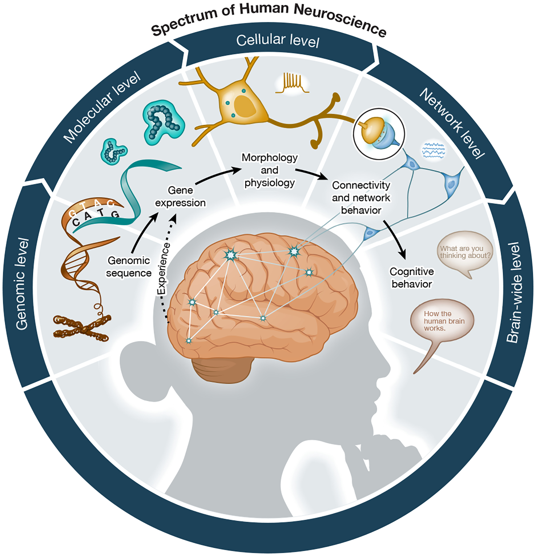
Cells in the human brain share the same genomic information but develop distinct patterns of gene expression. In turn, gene expression profiles correlate, and in many cases, regulate the abundance of proteins that build the cells and determine their distinct properties, such as morphology and physiology. Ensembles of neuronal cells give rise to synaptically-coupled neuronal networks that generate network-level properties. Coordinated activity of neurons in neural circuit give rise to motor output and cognition. Importantly, neuronal activity feeds back to control gene expression through activity-dependent molecular and cellular changes.
Ongoing efforts seek to characterize the diversity of cell types in the human brain using single cell or single nucleus transcriptomics33, chromatin state profiling, and epigenomics34–36. New and emerging efforts are beginning to extend the spectrum of methodologies to incorporate measurements of lipids, metabolites, proteins37, opening up new opportunities for deep molecular characterization of cell types in the human brain. It is important to recognize that many features of cell identity, such as the subcellular transcriptome, are not captured in those studies due to the limitations of current technologies38,39. We also must embrace the fact that a consensus definition of a cell type is currently lacking40, but even if the question of the absolute number of cell types in the brain cannot be fully addressed, we can be successful in defining their transcriptomic marker genes. The impact of census-scale transcriptomic studies have already led to the development of technologies that allow us to visualize transcriptomic cell types in tissue38,41–43 and begin to precisely localize them within a common coordinate framework44.
Linking cellular function and networks
Molecular cell states have primarily been assayed using postmortem human tissue specimens. However, the time needed to harvest and freeze postmortem tissue may result in significant changes in gene expression45. Furthermore, studies of postmortem tissue fail to capture the functional properties of cells, many of which could be fundamental to cell type definition (Figure 1). The extent to which transcriptomic cell types contribute to network dynamics is an open question, which is not even fully addressed in model organisms24,46. Thus, the advent of approaches that allow neurosurgical specimen to be maintained viable in culture for multiple days offers an alternative opportunity to study cell types within a functional microcircuit24,46,47 (Figure 2).
Figure 2: Central approaches to advance the study of human brain.
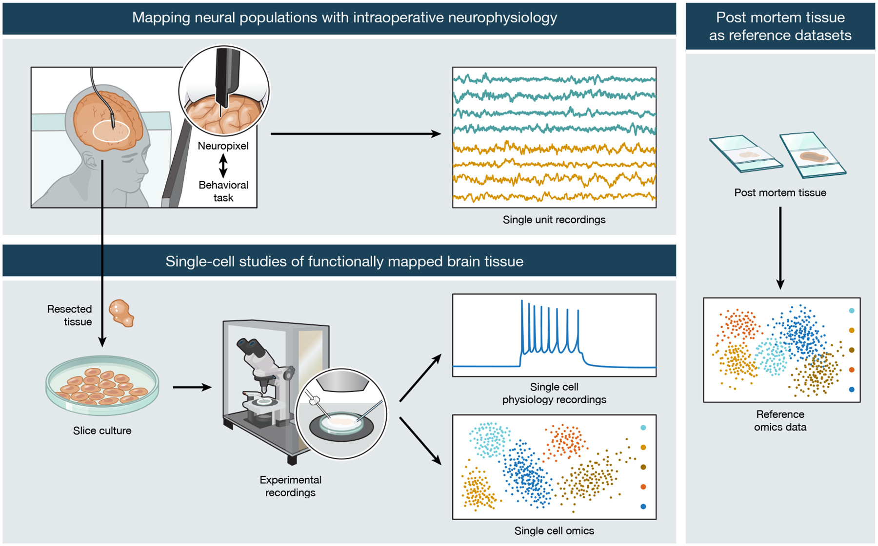
Intraoperative recordings on neural activity while patients are doing cognitive tasks can be conducted ethically and with patient consent to gain insight into neural correlates of human behavior (top left). Tissue resected from the brain can be maintained in ex vivo culture condition to facilitate single-cell electrophysiology and molecular profiling (bottom left). In parallel, postmortem tissue specimens provide generalizable insights across brain regions and broader patient population (right).
Human brain tissue resected from surgery has already begun to reveal new insights into how human neurons process information by leveraging human-specific neurophysiology such as unique dendritic compartmentalization and computation48–51. Since many of the epilepsy surgeries require the patients to be awake, ex vivo approaches also afford a rare opportunity of investigating the genetic and cellular composition of tissue that has been functionally mapped as the patient undergoes cognitive testing. For example, transcriptomic studies of resected tissue functionally mapped with episodic memory tasks have linked specific gene expression networks with oscillatory signatures of memory formation across multiple frequency bands52. Indeed, the ability of the ex vivo approach to dissect the cellular architecture of brain tissue whose networks have been functionally mapped by Neuropixels or ECOG during human cognitive tasks (i.e. language), is a key distinction between human neuroscience and other model systems.
Studying neurosurgically resected tissue in culture also provides a window to study subtype-specific synaptic connectivity maps, which will be critical in linking cell types to circuit and network function. Although advances in electron microscopy (EM) will soon enable whole mouse brain connectome studies53, achieving the scale necessary for human brain may be a lot more challenging. Moreover, strategies to visualize molecular cell types in EM workflows are less well developed. Virally introducing synaptically-connected molecular barcodes in ex vivo slices, and then profiling their connectivity with next-generation single-cell sequencing technologies, may enable us to overcome the limitations of cell-type identities and scale seen in EM connectome strategies54. Although ex vivo virally-mediated barcode tracing strategies inherently narrows study to the local microcircuit within each slice, the integration of cellular identity and single-cell transcriptomics with their synaptic connectivity maps may highlight intrinsic differences in how endogenous network activity motifs differ in disease states55–57.
To take a step further, we need to find efficient ways to relate in vivo cellular activity to cell type identity, such as developing approaches that can register the location of a cell during in vivo recording and determine its identity ex vivo. By preserving the location of the cells from in vivo recordings to their ex vivo positions, large multidimensional datasets that incorporate neurophysiology with single-cell sequencing techniques (transcriptomics, chromatin state profiling, and epigenomics) can be built. This can to some degree be accomplished using extracellular electrodes58. An alternative approach may be to leverage in silico models, which provide biorealistic approximations of brain function. Success of biorealistic in silico models trained with data from humans provide a representation, and predictions from the models can be tested as validation59,60.
Several issues need to be considered with the ex vivo tissue culture approach including 1) the generalizability of findings from tissue collected in epilepsy surgeries, 2) the additional technical variation of cell health in tissue culture, and 3) the constraints of studying a microcircuit in isolation. Since the majority of our tissue comes primarily from epilepsy patients, an important caveat is that conclusions from these studies may not routinely be generalizable to the broader patient population. However, this limitation does not necessarily discount the utility of insights derived from this patient cohort. At our institution, the majority of the patients undergo focused temporal lobectomies where the primary epileptogenic pathology is in subcortical structures such as the hippocampus and amygdala, and not necessarily in the cortex. Moreover, because experimental conditions are compared across epilepsy patients rather than control cohorts, other control methodologies can be used, such as matched post-mortem gene expression samples from epilepsy and control individuals52. In fact, the heterogeneity of epilepsy patients may itself pose a unique research opportunity since this patient population often harbors co-morbidities such as depression, anxiety, or autism, which may also be of interest. Furthermore, multiple non-neuronal tissue types such as endothelial cells of the cerebrovascular system may also be studied in tissue from epileptogenic patient population and be more broadly generalizable61. Similar considerations of generalizability would also apply for tissue collected during resections of deep brain tumors. Overall then, ex vivo human neuroscience approaches can be used to stimulate growth in the field and promote hypotheses which can be supported with other techniques including gene association, exome sequencing, or proteomic studies. Regarding technical variations introduced by tissue culture, neurons from cultured brain slices have been demonstrated to have stable electrophysiological properties over several weeks and are similar to acute slices as evidenced by patch clamp electrophysiology47,62,63. Finally, although the ex vivo approach results in microcircuits lacking its native in vivo inputs, we can still study questions of subtype composition, synaptic connectivity, cell-intrinsic physiology, and endogenous network motifs, which altogether can provide mechanistic insight into the larger intact network.
Lastly, human ex vivo tissue culture models can provide a number of unique opportunities to advance therapeutic opportunities. First, defining cellular states directly in disease tissue can reveal unexpected cellular, genetic, and intercellular phenotypes that could reveal unexpected disease transformations, changes that correlate with disease progression and inform cross-model studies of animal and in vitro systems61. The process of developing and validating new vectors for gene therapy such as cell-type specific enhancer discovery64 or AAV capsid engineering by directed evolution could be accelerated by incorporating human tissue-based experiments, thereby lowering the risks of such tools failing to translate from mice or monkey to human65.
Human neuroscience and clinical practice
Thus far, we have outlined a case for the need and the approaches to advance human neuroscience at the cellular and network levels, and how to connect those dimensions to typical brain function. While these advances yield exciting insights into what makes us human, they also highlight the unmet need to develop strategies to precisely modulate the neural network at the gene, cellular, or circuit level towards the benefit of neurological and psychiatric patients. This journey starts with the need to systematically define cellular function and dysfunction in the context of disease and, with the rapid gains in basic science knowledge, to develop new precision tools for cell- or circuit- specific treatments.
Genetic mapping and precision medicine.
Genomic, transcriptomic, and proteomic studies have defined a starting point to understand neuropathological mechanisms of many neurological and psychiatric conditions. Gene association and exome sequencing studies have begun to identify genetic risk architecture of disease such as with motor neuron disease66 and neurodegenerative diseases67,68, epilepsy69,70, autism spectrum disorders and neurodevelopmental disorders71,72, and schizophrenia73,74. Thanks to the rapid advances in sequencing technologies, genetic diagnosis75 is being applied to improve diagnostic capabilities. Identifying variants in risk genes for complex conditions such as Autism Spectrum Disorder and Epilepsy have been increasingly incorporated in clinical work-up76,77 and can provide guidance when symptoms point to a suspected diagnosis.
However, genomic variants identified at the plasma level explain a small fraction of cases in many neurological and psychiatric cases78. The ability to access human brain tissue, such as via neurosurgical resections, has revealed a unique set of mutations that emerge outside the germline and led to a growing appreciation for somatic mosaicism in the role of typical brain development and in neurological disease79–82. Mutations in this scenario emerge later in development after the zygote state, leading to organ tissues that are mosaic, with only a subset of cells containing the mutation. Such somatic mutations have specifically been observed in tissue obtained from neurosurgical patients, such as those with focal epilepsy who have undergone resection to remove a lesion, such as in cortical dysplasia or hemimegalencephaly83,84. This has led to the identification of new disease risk genes for focal cortical dysplasias and HME, including AKT385 and DEPDC586, and a converging role of the mTOR pathway in neurodevelopmental-associated epilepsy87. These findings would not have been possible without the interrogation of human neurosurgical material.
Gain- or loss-of-function of genes identified in these studies increases the risk for a particular condition, and this information can guide pharmaceutical development, or even repurposing of old drugs. For example, Dravet Syndrome is caused by loss of function mutations in a gene SCN1A, which encodes a sodium channel. Identification of the causal gene has led to practical ways to improve patient outcomes including better treatment strategy (avoiding certain seizure medications) and use of novel pharmacologic agents, including fenfluramine and cannabidiol88.
Furthermore, mutational burden information can be integrated with cell type-specific gene expression information profiles to highlight patterns of select cell type vulnerability to disease-causing mutations. One example of this is the identification of TREM2 as a genetic risk factor for Alzheimer’s Disease89. TREM2 is selectively expressed in microglia90, a finding which has sparked substantial efforts to understand the role of the innate immune system in Alzheimer’s Disease. Of course, gene expression in any one cell type may not be necessary or sufficient to explain the full spectrum of disease symptoms, but identifying profiles of gene expression may represent an important step towards systematic interrogation of cell type specific contributions patient specific mutations to disease processes. Such information can also guide efforts to develop improved cellular models that could serve as platforms to interrogate disease mechanisms and serve as preclinical models.
Although vast majority of single cell reference atlases of human cell type diversity do not extensively characterize mRNA isoform, an important yet underestimated ramification of cell type specific atlases is the potential to identify pathogenic and cell type specific mRNA isoforms. Antisense oligonucleotides (ASOs) are single-stranded synthetic short molecules that bind to target mRNA and lead to target specific knockdown. ASOs have been used for treatment of Duchenne muscular dystrophy91,92 and spinal muscular atrophy93. The biggest limitations to the use of ASOs are ensuring correct sequence matching to target genes (to optimize gene targeting of all isoforms and prevent off-target effects) and delivery, given these molecules are short-lived and cannot cross the blood-brain barrier. Isoform-resolved single cell transcriptomic atlases could accelerate the identification of potential targets for this emerging class of therapeutic tools.
The ability to integrate the increasing datasets available94–97 into machine-learning enabled formats98 will enable deep learning approaches to combined the genetic, clinical and environmental factors into a more cohesive framework to create tailored and accurate therapeutic strategies.
Cell-type specific molecular access
Emergence of new tools for cell type-specific molecular access to monitor, target and manipulate specific cell types in the human brain open up opportunities to pursue mechanistic and therapeutic studies. Gene therapy offers a direct way to repair or replace a defective gene, an approach that is highly suited for monogenic conditions. Adeno-associated viral (AAV) vectors represent an approach that has been successfully applied to target nervous system tissue99,100. AAV vectors can infect many cell types, show low immunogenicity, and enable long-term expression of introduced transgene without requiring host chromosome integration. One successful example in neurology is spinal muscular atrophy, a devastating neurodegenerative neuromuscular disease101 caused by mutations in SMN1 or SMN2102. The positive results from the initial cohort has led to enthusiasm about the potential for in vivo gene therapy, such as via AAV vectors, and has served as a guide for gene therapy trials in other conditions103. There are currently trials to treat conditions such as Canavan disease (with mutations in ASPA that makes aspartoacylase), Batten disease (a lysosomal storage disorder due to the loss of tripeptidyl peptidase 1), and aromatic-L-amino acid decarboxylase (AADC) deficiency104,105. However, most AAVs used in current clinical trials have very broad tropism, and as we begin to better understand neurological disease at the cell type and circuit level, it will become increasingly important to develop cell type specific AAVs. Advances in the engineering of AAV capsids and identification of cell-type specific enhancer elements will allow for more precise AAV vector targeting106–108 and applicability across species64. These next generation of vectors will allow for the delivery of a variety of effector molecules not limited to causal disease genes, but will be increasingly tailored to cargos such as optogenetic or chemogenetic actuators of neural activity.
Ethics and data in human neuroscience
Human neuroscience research depends critically on patient volunteers. Participation should involve informed consent, which describes both the risks for tissue collection or neural device implantation, and also how the data generated can be used and shared with the research community. This is especially important since the immediate benefit from surgical patients participating as research subjects are currently limited and the risks are potentially severe, in both obvious and unpredictable ways. For tissue collection, it is paramount patients understand that participation in tissue collection does not change their surgical plan. Only tissue that is clinically indicated for resection are collected for research. The ethical frameworks for human research with neural devices have been reviewed extensively by the NIH BRAIN Initiative Neuroethics Working Group and discusses the analysis of risk, informed consent, and posttrial responsibilities to research participants undergoing implantation of neural devices109. With regards specifically to data sharing, the Working Group state that the social and scientific value of data sharing should be weighed against the robust analyses of privacy risks, and that sharing data may be a key posttrial responsibility. In practice, many types of human neurophysiology data are intrinsically difficult to de-identify, particularly the high-dimensional datasets obtained from recording technologies described earlier in this perspective. However, informed consent should recognize that unforeseen advances in machine-learning (ML) and artificial intelligence (AI) may risk patient de-identification, particularly as neurophysiology data begin to be superimposed by the patient’s corresponding transcriptomics, epigenomics, and proteomics datasets. The merging and analyses of this complex dataset will require the computing and analytical power of ML and AI. In this context then, what constitutes as informed consent will need to be revisited as we increasingly rely on these technologies, and the degree to which their conclusions arise from a spectrum of “white-box” or “black-box” approaches (i.e. explainable vs unexplainable models)110. In the near future, an important discussion for all stakeholders is what safeguards to have in place as AI advances towards artificial general intelligence111. One way to maintain faith in the informed consent process is for the academic research community to make transparent any partnerships between academics and industry technology companies, particularly since private industries are significant drivers of AI advances.
Recently, Research Opportunities in Humans (ROH) Consortium has developed a series of best practices around engaging patients in research to maintain the integrity of clinical care and ensure voluntariness of participation112. However, further discussions are needed especially around data sharing practices. Considerations around the privacy and ownership of the research data have been quite advanced in other fields such as genomics, where public benefits to datasets involving large numbers of study participants have been quite clear. What are the ramifications for broad dissemination of data? Who should have access to the data? What are the public benefits and what are the risks? Answers to those questions are less clearly defined in neuroscience compared to genomics.
Sharing research findings based on data derived from study participants is generally viewed as safe because the raw data is de-identified, or that the underlying raw data did not carry information that would pose a possibility of re-identifying the study participants. New data sharing policies that set FAIR (findability, accessibility, interoperability, and reusability) principles at the centerpiece of federally funded research are set to revolutionize biomedical research113, but also prompt a reflection upon the risks of data sharing. Even when data is de-identified, with the advent of very large-scale datasets, the possibility of re-identification becomes a point of concern. Nowhere is this more clear than in the area of genomics, where naturally occurring human genetic variation can be used with some degree of confidence to re-identify an individual114, which is particularly important as commercial providers of genome sequencing create incentives to share genomic material of large fragment of the population115. To account for the possibility of re-identifying study participants, consent forms now often implement explicit language informing the participants about the potential risk that the data generated as a result of their participation could be used to re-identify them, such that an individual signing a consent form is aware of the ramifications. However, as discussed above it is difficult to fully predict how future technologies or analytical approaches might open up new avenues to use the data. To limit the risk of potential misuse of such information, researchers can make study data available through controlled access archives, which impose restrictions on how the data can be utilized. At this point in time, these nuanced considerations have been largely limited to genomic data because of the clear evidence that genetic sequence information is unique to every individual and have implications to clinical management.116,117.
In genomics, population-scale studies have demonstrated the importance of sequence-level variation as it relates to identifying individuals with genetic risk who could be targeted for screening and early detection, which in the case of cancer mutations can substantially increase survival118. In neuropsychiatric disease, recent investigations in somatic copy-number variants have detected potential genetic loci in increasing schizophrenia risk119. In terms of affecting medical management, multiple examples exist where information of a patient’s genetic background can lead to a more precise treatment plan, such as changes in clopidogrel metabolism based on CYP2C19 genotype120 and increasing progression-free survival using targeted IDH inhibitors in IDH-mutant glioma121. Thus, genomics information has been transformative thanks to two properties: genetic sequence information is unique to every individual, and such information has clear implications for disease risk assessment, prevention, and treatment strategy. Another revolution poised to take advantage of genomic variant detection includes recent developments in CRISPR therapeutics122.
While neuroscience data has currently no clear equivalents of these two properties, ongoing large-scale data generation efforts will begin to test this assumption. Single cell genomics datasets are already beginning to uncover molecular states of cells associated with diseases such as Alzheimer’s Disease123–125. Advent of systematic taxonomies of cell types in the human brain will provide a quantitative structure to systematically compare these cellular phenotypes across disorders126, and the knowledge generated as a result of such comparisons will likely lead to new therapeutic interventions that precisely target the underlying cells and circuits. Although the full potential of single cell atlases has not been fully demonstrated, there are multiple ways in which such data will likely contribute to addressing treatment needs in neurological disorders, as discussed above.
What is less apparent is how data collected during intraoperative neural recordings should be managed. It is clear that neural activity data carries important information about the underlying disease states, and large-scale datasets collected across hundreds of individuals will likely reveal common patterns of neural function that could provide inroads into personalized therapy. In this scenario data sharing would ultimately benefit individual patients. However, as transcriptomic and epigenomic information are superimposed onto the physiology datasets, human neuroscience may begin to uncover the genetic and physiological codes that underlie our cognitive functions on personal and population levels. The research and broader community will need to be prepared about how to explore these questions since there is real potential for group-level harm and using data to affirm existing biases against certain groups of people. It will require the neuroscience community to engage not just patients, but the general public about the ramifications for broad dissemination of data, the public benefits and risks to different degrees of data access, and the perspectives of different types of communities (race, ethnicity, faith, etc) on how to conduct and apply the findings from human neuroscience research. At first glance, it is sensible that the data sharing of neural activity would be well served to follow similar principles to genomic information. This approach ensures that all findable, accessible, interoperable, and resuable datasets collected from an individual could be removed if requested by the patient. However, patient and community engagement will be just as critical to support a diversification of neuroscience research participants which is required for understanding of human brain function that is inclusive of all people.
The success of human neuroscience
Together, understanding fundamental principles of human brain structure and function, mechanistic interrogation of genetic, cellular and neurophysiological properties of neurons and glia, and the development of tools to precisely monitor, target and replace cells of the brain, we will begin to deliver on the ambitious goals of the BRAIN initiative. But how can we best capitalize on these discoveries and technological advances? What is the future of human neuroscience now that we begin to gain the right tools and mechanisms to study the human brain, and how should we do this responsibly and ethically?
One of the most transformative lessons from the human genome project has been that in order to enable rapid data and knowledge sharing, the scientific community needs to adopt the use of accessible data sharing platforms and data browsers, which organize data in a common reference. Similar efforts are beginning to organize around proposals for cellular taxonomies126, but at the current pace of data production, we also predict an even more pressing need to accelerate efforts for adoption of cloud-enabled data repositories127, adoption of tools for integrative analysis and annotation, including large language models. Focus of such analyses must include not only the definition of cell types and states but also phenotypes, as opposed to reference taxonomies, as well as development of comparable tools for epigenetic datasets that compare chromatin accessibility. Non-discrete, continuous sources of variation related to cellular trajectories that may exist during development or state transition may pose significant analytical challenges. Responsible data sharing and use policies for genetic variant information must be considered128. There is an emerging but unmet need to develop standards of data sharing in neurophysiology that could serve as a foundation to accelerate secure data platforms for analysis of human neurophysiological datasets derived from surgically implanted probes, especially at the time when commercial entities begin to consider adoption of such technologies.
The second key issue is that an approach to science will have to fundamentally change from single investigator driven projects to community-driven efforts, as evidenced by similar collaborative human neuroscience efforts to ours across the United States (i.e. Massachusetts General Hospital, Stanford, Baylor, Southwestern, Cedar-Sinai). This will bring several major benefits. Partnerships involving basic science researchers, computational scientists, physician scientists, and physicians will galvanize projects that interrogate questions in human neuroscience across scales, and across models. We argue that such partnerships should include engagement efforts of communities of interest, including patient groups and study participants who ultimately consent to share their information, data, and tissue information with the trust that such data will be used in a responsible manner, and which could ultimately lead to a better understanding of neurological conditions and neurodiversity, and to development of tools to modulate neural functions. We believe that a revolution in biomedical science has already begun, as evidenced by the formation of BRAIN Initiative Network efforts129 to tackle similar challenges in mice and non-human primates, and to begin to set the stage to address these questions in human.
Finally, for all the advances to happen efficiently and equitably, patient volunteers and their families must be invited to the table. Scientists and physician-scientists can ensure respectful and affirming community engagement through partnership with patient advocacy groups, meeting with community partners, and offering accessibility through outreach and educational events130–132. These practices require preparation of team leaders and members and getting the appropriate skills to perform these tasks. Engagement involves creating trust, allowing for dynamic communication, and ultimately giving back to the community, through the discovery and increased awareness of the neuroscience and neuropathology133. Healing happens with the potential for therapy but also with the acknowledgement and sharing of gained knowledge.
We propose that the era of human neuroscience has begun and will lead to transformative discoveries in fundamental and clinical neuroscience over the next decade, and critical to its success is the ability to build a Human Neuroscience community that embraces the principles of data sharing that have enabled the genomics community to succeed.
Footnotes
Competing interests
The authors declare no competing interests.
References
- 1.Burns SP, Xing D & Shapley RM Comparisons of the dynamics of local field potential and multiunit activity signals in macaque visual cortex. J Neurosci 30, 13739–13749 (2010). [DOI] [PMC free article] [PubMed] [Google Scholar]
- 2.Ahmadi N, Constandinou TG & Bouganis CS Inferring entire spiking activity from local field potentials. Sci Rep 11, 19045 (2021). [DOI] [PMC free article] [PubMed] [Google Scholar]
- 3.Tseng HA & Han X Distinct Spiking Patterns of Excitatory and Inhibitory Neurons and LFP Oscillations in Prefrontal Cortex During Sensory Discrimination. Front Physiol 12, 618307 (2021). [DOI] [PMC free article] [PubMed] [Google Scholar]
- 4.Cho KK et al. Gamma rhythms link prefrontal interneuron dysfunction with cognitive inflexibility in Dlx5/6(+/−) mice. Neuron 85, 1332–1343 (2015). [DOI] [PMC free article] [PubMed] [Google Scholar]
- 5.Helfrich RF & Knight RT Oscillatory Dynamics of Prefrontal Cognitive Control. Trends Cogn Sci 20, 916–930 (2016). [DOI] [PMC free article] [PubMed] [Google Scholar]
- 6.Kamigaki T & Dan Y Delay activity of specific prefrontal interneuron subtypes modulates memory-guided behavior. Nat Neurosci 20, 854–863 (2017). [DOI] [PMC free article] [PubMed] [Google Scholar]
- 7.Pinto L & Dan Y Cell-Type-Specific Activity in Prefrontal Cortex during Goal-Directed Behavior. Neuron 87, 437–450 (2015). [DOI] [PMC free article] [PubMed] [Google Scholar]
- 8.Lagler M et al. Divisions of Identified Parvalbumin-Expressing Basket Cells during Working Memory-Guided Decision Making. Neuron 91, 1390–1401 (2016). [DOI] [PubMed] [Google Scholar]
- 9.Kvitsiani D et al. Distinct behavioural and network correlates of two interneuron types in prefrontal cortex. Nature 498, 363–366 (2013). [DOI] [PMC free article] [PubMed] [Google Scholar]
- 10.Liebe S, Hoerzer GM, Logothetis NK & Rainer G Theta coupling between V4 and prefrontal cortex predicts visual short-term memory performance. Nat Neurosci 15, 456–62, S1 (2012). [DOI] [PubMed] [Google Scholar]
- 11.Buschman TJ, Denovellis EL, Diogo C, Bullock D & Miller EK Synchronous oscillatory neural ensembles for rules in the prefrontal cortex. Neuron 76, 838–846 (2012). [DOI] [PMC free article] [PubMed] [Google Scholar]
- 12.Hsieh HL, Wong YT, Pesaran B & Shanechi MM Multiscale modeling and decoding algorithms for spike-field activity. J Neural Eng 16, 016018 (2019). [DOI] [PubMed] [Google Scholar]
- 13.Fu Z et al. The geometry of domain-general performance monitoring in the human medial frontal cortex. Science 376, eabm9922 (2022). [DOI] [PMC free article] [PubMed] [Google Scholar]; This study is an example of using Behnke-Fried electrodes to collect single neuron activity in large patient cohorts.
- 14.Steinmetz NA, Koch C, Harris KD & Carandini M Challenges and opportunities for large-scale electrophysiology with Neuropixels probes. Curr Opin Neurobiol 50, 92–100 (2018). [DOI] [PMC free article] [PubMed] [Google Scholar]
- 15.Chung JE et al. High-density single-unit human cortical recordings using the Neuropixels probe. Neuron 110, 2409–2421.e3 (2022). [DOI] [PubMed] [Google Scholar]; This study and Paulk et al are one of first to demonstrate use of neuropixels in human cortex
- 16.Khanna AR et al. Single-neuronal elements of speech production in humans. Nature 626, 603–610 (2024). [DOI] [PMC free article] [PubMed] [Google Scholar]
- 17.Leonard MK et al. Large-scale single-neuron speech sound encoding across the depth of human cortex. Nature (2023). [DOI] [PMC free article] [PubMed] [Google Scholar]; This study highlights use of Neuropixels for investigating columnar processing in human speech encoding
- 18.Trautmann EM et al. Large-scale high-density brain-wide neural recording in nonhuman primates. bioRxiv 2023.02.01.526664 (2023). [DOI] [PMC free article] [PubMed] [Google Scholar]
- 19.Tischbirek CH et al. In Vivo Functional Mapping of a Cortical Column at Single-Neuron Resolution. Cell Rep 27, 1319–1326.e5 (2019). [DOI] [PubMed] [Google Scholar]
- 20.Khodagholy D et al. NeuroGrid: recording action potentials from the surface of the brain. Nat Neurosci 18, 310–315 (2015). [DOI] [PMC free article] [PubMed] [Google Scholar]
- 21.Tchoe Y et al. Human brain mapping with multithousand-channel PtNRGrids resolves spatiotemporal dynamics. Sci Transl Med 14, eabj1441 (2022). [DOI] [PMC free article] [PubMed] [Google Scholar]
- 22.Paulk AC et al. Microscale Physiological Events on the Human Cortical Surface. Cereb Cortex 31, 3678–3700 (2021). [DOI] [PMC free article] [PubMed] [Google Scholar]
- 23.Paulk AC et al. Large-scale neural recordings with single neuron resolution using Neuropixels probes in human cortex. Nat Neurosci 25, 252–263 (2022). [DOI] [PubMed] [Google Scholar]; This study and Chung et al are one of first to demonstrate use of neuropixels in human cortex
- 24.Bugeon S et al. A transcriptomic axis predicts state modulation of cortical interneurons. Nature 607, 330–338 (2022). [DOI] [PMC free article] [PubMed] [Google Scholar]
- 25.Pollen AA et al. Low-coverage single-cell mRNA sequencing reveals cellular heterogeneity and activated signaling pathways in developing cerebral cortex. Nat Biotechnol 32, 1053–1058 (2014). [DOI] [PMC free article] [PubMed] [Google Scholar]
- 26.Nowakowski TJ et al. Spatiotemporal gene expression trajectories reveal developmental hierarchies of the human cortex. Science 358, 1318–1323 (2017). [DOI] [PMC free article] [PubMed] [Google Scholar]; This study applies next-generation single-cell RNA sequencing technologies to profile human cortical cell atlases in brain development
- 27.Darmanis S et al. A survey of human brain transcriptome diversity at the single cell level. Proc Natl Acad Sci U S A 112, 7285–7290 (2015). [DOI] [PMC free article] [PubMed] [Google Scholar]
- 28.Macosko EZ et al. Highly Parallel Genome-wide Expression Profiling of Individual Cells Using Nanoliter Droplets. Cell 161, 1202–1214 (2015). [DOI] [PMC free article] [PubMed] [Google Scholar]; This study introduces a key method in development of next-generation single-cell RNA sequencing technologies
- 29.Langlieb J et al. The cell type composition of the adult mouse brain revealed by single cell and spatial genomics. bioRxiv 2023.03.06.531307 (2023). [Google Scholar]
- 30.Yao Z et al. A high-resolution transcriptomic and spatial atlas of cell types in the whole mouse brain. Nature 624, 317–332 (2023). [DOI] [PMC free article] [PubMed] [Google Scholar]; This study uses single-cell RNA sequencing and spatial transcriptomics to atlas the entire mouse brain.
- 31.Siletti K et al. Transcriptomic diversity of cell types across the adult human brain. Science 382, eadd7046 (2023). [DOI] [PubMed] [Google Scholar]; This study is an initial draft of a cell atlas of the human brain.
- 32.Clark IC et al. Microfluidics-free single-cell genomics with templated emulsification. Nat Biotechnol 41, 1557–1566 (2023). [DOI] [PMC free article] [PubMed] [Google Scholar]
- 33.Bakken TE et al. Comparative cellular analysis of motor cortex in human, marmoset and mouse. Nature 598, 111–119 (2021). [DOI] [PMC free article] [PubMed] [Google Scholar]; This study is an example that highlights unique cell type-specific transcriptomic and physiologic properties of human neurons compared to model systems.
- 34.Luo C et al. Single-cell methylomes identify neuronal subtypes and regulatory elements in mammalian cortex. Science 357, 600–604 (2017). [DOI] [PMC free article] [PubMed] [Google Scholar]
- 35.Ziffra RS et al. Single-cell epigenomics reveals mechanisms of human cortical development. Nature 598, 205–213 (2021). [DOI] [PMC free article] [PubMed] [Google Scholar]
- 36.Luo C et al. Single nucleus multi-omics identifies human cortical cell regulatory genome diversity. Cell Genom 2, 100107 (2022). [DOI] [PMC free article] [PubMed] [Google Scholar]
- 37.Gatto L et al. Initial recommendations for performing, benchmarking and reporting single-cell proteomics experiments. Nat Methods 20, 375–386 (2023). [DOI] [PMC free article] [PubMed] [Google Scholar]
- 38.Alon S et al. Expansion sequencing: Spatially precise in situ transcriptomics in intact biological systems. Science 371, eaax2656 (2021). [DOI] [PMC free article] [PubMed] [Google Scholar]
- 39.Niu M et al. Droplet-based transcriptome profiling of individual synapses. Nat Biotechnol 41, 1332–1344 (2023). [DOI] [PubMed] [Google Scholar]
- 40.Zeng H What is a cell type and how to define it. Cell 185, 2739–2755 (2022). [DOI] [PMC free article] [PubMed] [Google Scholar]
- 41.Fang R et al. Conservation and divergence of cortical cell organization in human and mouse revealed by MERFISH. Science 377, 56–62 (2022). [DOI] [PMC free article] [PubMed] [Google Scholar]
- 42.Zhang D et al. Spatial epigenome-transcriptome co-profiling of mammalian tissues. Nature 616, 113–122 (2023). [DOI] [PMC free article] [PubMed] [Google Scholar]
- 43.Wang X et al. Three-dimensional intact-tissue sequencing of single-cell transcriptional states. Science 361, eaat5691 (2018). [DOI] [PMC free article] [PubMed] [Google Scholar]
- 44.Glasser MF et al. A multi-modal parcellation of human cerebral cortex. Nature 536, 171–178 (2016). [DOI] [PMC free article] [PubMed] [Google Scholar]
- 45.Liharska LE et al. A study of gene expression in the living human brain. medRxiv 2023.04.21.23288916 (2023). [Google Scholar]
- 46.Schneider A et al. Transcriptomic cell type structures in vivo neuronal activity across multiple timescales. Cell Rep 42, 112318 (2023). [DOI] [PMC free article] [PubMed] [Google Scholar]
- 47.Ting JT et al. A robust ex vivo experimental platform for molecular-genetic dissection of adult human neocortical cell types and circuits. Sci Rep 8, 8407 (2018). [DOI] [PMC free article] [PubMed] [Google Scholar]
- 48.Beaulieu-Laroche L et al. Enhanced Dendritic Compartmentalization in Human Cortical Neurons. Cell 175, 643–651.e14 (2018). [DOI] [PMC free article] [PubMed] [Google Scholar]
- 49.Moradi Chameh H et al. Diversity amongst human cortical pyramidal neurons revealed via their sag currents and frequency preferences. Nat Commun 12, 2497 (2021). [DOI] [PMC free article] [PubMed] [Google Scholar]
- 50.Kalmbach BE et al. h-Channels Contribute to Divergent Intrinsic Membrane Properties of Supragranular Pyramidal Neurons in Human versus Mouse Cerebral Cortex. Neuron 100, 1194–1208.e5 (2018). [DOI] [PMC free article] [PubMed] [Google Scholar]
- 51.Beaulieu-Laroche L et al. Allometric rules for mammalian cortical layer 5 neuron biophysics. Nature 600, 274–278 (2021). [DOI] [PMC free article] [PubMed] [Google Scholar]
- 52.Berto S et al. Gene-expression correlates of the oscillatory signatures supporting human episodic memory encoding. Nat Neurosci 24, 554–564 (2021). [DOI] [PMC free article] [PubMed] [Google Scholar]
- 53.Mahalingam G et al. A scalable and modular automated pipeline for stitching of large electron microscopy datasets. Elife 11, e76534 (2022). [DOI] [PMC free article] [PubMed] [Google Scholar]
- 54.Chen X et al. High-Throughput Mapping of Long-Range Neuronal Projection Using In Situ Sequencing. Cell 179, 772–786.e19 (2019). [DOI] [PMC free article] [PubMed] [Google Scholar]
- 55.Foss-Feig JH et al. Searching for Cross-Diagnostic Convergence: Neural Mechanisms Governing Excitation and Inhibition Balance in Schizophrenia and Autism Spectrum Disorders. Biol Psychiatry 81, 848–861 (2017). [DOI] [PMC free article] [PubMed] [Google Scholar]
- 56.Krystal JH et al. Impaired Tuning of Neural Ensembles and the Pathophysiology of Schizophrenia: A Translational and Computational Neuroscience Perspective. Biol Psychiatry 81, 874–885 (2017). [DOI] [PMC free article] [PubMed] [Google Scholar]
- 57.Opris I & Casanova MF Prefrontal cortical minicolumn: from executive control to disrupted cognitive processing. Brain 137, 1863–1875 (2014). [DOI] [PMC free article] [PubMed] [Google Scholar]
- 58.Li Q et al. Multimodal charting of molecular and functional cell states via in situ electro-sequencing. Cell 186, 2002–2017.e21 (2023). [DOI] [PMC free article] [PubMed] [Google Scholar]
- 59.Billeh YN et al. Systematic Integration of Structural and Functional Data into Multi-scale Models of Mouse Primary Visual Cortex. Neuron 106, 388–403.e18 (2020). [DOI] [PubMed] [Google Scholar]
- 60.Buchin A et al. Multi-modal characterization and simulation of human epileptic circuitry. Cell Rep 41, 111873 (2022). [DOI] [PMC free article] [PubMed] [Google Scholar]
- 61.Winkler EA et al. A single-cell atlas of the normal and malformed human brain vasculature. Science 375, eabi7377 (2022). [DOI] [PMC free article] [PubMed] [Google Scholar]; This study is an example of how ex vivo platform can be utilized to study non-neuronal cell types in human brain and disease.
- 62.Eugène E et al. An organotypic brain slice preparation from adult patients with temporal lobe epilepsy. J Neurosci Methods 235, 234–244 (2014). [DOI] [PMC free article] [PubMed] [Google Scholar]
- 63.Schwarz N et al. Long-term adult human brain slice cultures as a model system to study human CNS circuitry and disease. Elife 8, (2019). [DOI] [PMC free article] [PubMed] [Google Scholar]
- 64.Mich JK et al. Functional enhancer elements drive subclass-selective expression from mouse to primate neocortex. Cell Rep 34, 108754 (2021). [DOI] [PMC free article] [PubMed] [Google Scholar]
- 65.Zhu D et al. Optimal trade-off control in machine learning-based library design, with application to adeno-associated virus (AAV) for gene therapy. bioRxiv (2021). [DOI] [PMC free article] [PubMed] [Google Scholar]
- 66.Mercuri E, Pera MC, Scoto M, Finkel R & Muntoni F Spinal muscular atrophy - insights and challenges in the treatment era. Nat Rev Neurol 16, 706–715 (2020). [DOI] [PubMed] [Google Scholar]
- 67.Neuner SM, Tcw J & Goate AM Genetic architecture of Alzheimer’s disease. Neurobiol Dis 143, 104976 (2020). [DOI] [PMC free article] [PubMed] [Google Scholar]
- 68.Tabrizi SJ, Flower MD, Ross CA & Wild EJ Huntington disease: new insights into molecular pathogenesis and therapeutic opportunities. Nat Rev Neurol 16, 529–546 (2020). [DOI] [PubMed] [Google Scholar]
- 69.Niestroj LM et al. Epilepsy subtype-specific copy number burden observed in a genome-wide study of 17 458 subjects. Brain 143, 2106–2118 (2020). [DOI] [PMC free article] [PubMed] [Google Scholar]
- 70.Epi4K, C. et al. De novo mutations in epileptic encephalopathies. Nature 501, 217–221 (2013). [DOI] [PMC free article] [PubMed] [Google Scholar]
- 71.Wang T et al. Integrated gene analyses of de novo variants from 46,612 trios with autism and developmental disorders. Proc Natl Acad Sci U S A 119, e2203491119 (2022). [DOI] [PMC free article] [PubMed] [Google Scholar]
- 72.Fu JM et al. Rare coding variation provides insight into the genetic architecture and phenotypic context of autism. Nat Genet 54, 1320–1331 (2022). [DOI] [PMC free article] [PubMed] [Google Scholar]
- 73.Singh T et al. Rare coding variants in ten genes confer substantial risk for schizophrenia. Nature 604, 509–516 (2022). [DOI] [PMC free article] [PubMed] [Google Scholar]
- 74.Liu D et al. Schizophrenia risk conferred by rare protein-truncating variants is conserved across diverse human populations. Nat Genet 55, 369–376 (2023). [DOI] [PMC free article] [PubMed] [Google Scholar]
- 75.Lin X et al. Integrating Genetic Structural Variations and Whole-Genome Sequencing Into Clinical Neurology. Neurol Genet 8, e200005 (2022). [DOI] [PMC free article] [PubMed] [Google Scholar]
- 76.Neale BM et al. Patterns and rates of exonic de novo mutations in autism spectrum disorders. Nature 485, 242–245 (2012). [DOI] [PMC free article] [PubMed] [Google Scholar]
- 77.Mitra I et al. Patterns of de novo tandem repeat mutations and their role in autism. Nature 589, 246–250 (2021). [DOI] [PMC free article] [PubMed] [Google Scholar]
- 78.Nishioka M, Bundo M, Iwamoto K & Kato T Somatic mutations in the human brain: implications for psychiatric research. Mol Psychiatry 24, 839–856 (2019). [DOI] [PMC free article] [PubMed] [Google Scholar]
- 79.Rehen SK et al. Constitutional aneuploidy in the normal human brain. J Neurosci 25, 2176–2180 (2005). [DOI] [PMC free article] [PubMed] [Google Scholar]
- 80.Wang Y et al. Comprehensive identification of somatic nucleotide variants in human brain tissue. Genome Biol 22, 92 (2021). [DOI] [PMC free article] [PubMed] [Google Scholar]
- 81.Luquette LJ et al. Single-cell genome sequencing of human neurons identifies somatic point mutation and indel enrichment in regulatory elements. Nat Genet 54, 1564–1571 (2022). [DOI] [PMC free article] [PubMed] [Google Scholar]
- 82.Chung C et al. Comprehensive multi-omic profiling of somatic mutations in malformations of cortical development. Nat Genet 55, 209–220 (2023). [DOI] [PMC free article] [PubMed] [Google Scholar]
- 83.Hua Y & Crino PB Single cell lineage analysis in human focal cortical dysplasia. Cereb Cortex 13, 693–699 (2003). [DOI] [PubMed] [Google Scholar]
- 84.Rivière JB et al. De novo germline and postzygotic mutations in AKT3, PIK3R2 and PIK3CA cause a spectrum of related megalencephaly syndromes. Nat Genet 44, 934–940 (2012). [DOI] [PMC free article] [PubMed] [Google Scholar]
- 85.Poduri A et al. Somatic activation of AKT3 causes hemispheric developmental brain malformations. Neuron 74, 41–48 (2012). [DOI] [PMC free article] [PubMed] [Google Scholar]
- 86.Ribierre T et al. Second-hit mosaic mutation in mTORC1 repressor DEPDC5 causes focal cortical dysplasia-associated epilepsy. J Clin Invest 128, 2452–2458 (2018). [DOI] [PMC free article] [PubMed] [Google Scholar]
- 87.Baldassari S et al. Dissecting the genetic basis of focal cortical dysplasia: a large cohort study. Acta Neuropathol 138, 885–900 (2019). [DOI] [PMC free article] [PubMed] [Google Scholar]
- 88.Wirrell EC et al. Optimizing the Diagnosis and Management of Dravet Syndrome: Recommendations From a North American Consensus Panel. Pediatr Neurol 68, 18–34.e3 (2017). [DOI] [PubMed] [Google Scholar]
- 89.Gratuze M, Leyns CEG & Holtzman DM New insights into the role of TREM2 in Alzheimer’s disease. Mol Neurodegener 13, 66 (2018). [DOI] [PMC free article] [PubMed] [Google Scholar]
- 90.Ulland TK & Colonna M TREM2 - a key player in microglial biology and Alzheimer disease. Nat Rev Neurol 14, 667–675 (2018). [DOI] [PubMed] [Google Scholar]
- 91.Braasch DA, Liu Y & Corey DR Antisense inhibition of gene expression in cells by oligonucleotides incorporating locked nucleic acids: effect of mRNA target sequence and chimera design. Nucleic Acids Res 30, 5160–5167 (2002). [DOI] [PMC free article] [PubMed] [Google Scholar]
- 92.Bladen CL et al. The TREAT-NMD DMD Global Database: analysis of more than 7,000 Duchenne muscular dystrophy mutations. Hum Mutat 36, 395–402 (2015). [DOI] [PMC free article] [PubMed] [Google Scholar]
- 93.Finkel RS et al. Treatment of infantile-onset spinal muscular atrophy with nusinersen: a phase 2, open-label, dose-escalation study. Lancet 388, 3017–3026 (2016). [DOI] [PubMed] [Google Scholar]
- 94.Hampel H et al. The foundation and architecture of precision medicine in neurology and psychiatry. Trends Neurosci 46, 176–198 (2023). [DOI] [PMC free article] [PubMed] [Google Scholar]
- 95.Knowles JK et al. Precision medicine for genetic epilepsy on the horizon: Recent advances, present challenges, and suggestions for continued progress. Epilepsia 63, 2461–2475 (2022). [DOI] [PMC free article] [PubMed] [Google Scholar]
- 96.Varma VR et al. Brain and blood metabolite signatures of pathology and progression in Alzheimer disease: A targeted metabolomics study. PLoS Med 15, e1002482 (2018). [DOI] [PMC free article] [PubMed] [Google Scholar]
- 97.Noori A, Mezlini AM, Hyman BT, Serrano-Pozo A & Das S Systematic review and meta-analysis of human transcriptomics reveals neuroinflammation, deficient energy metabolism, and proteostasis failure across neurodegeneration. Neurobiol Dis 149, 105225 (2021). [DOI] [PMC free article] [PubMed] [Google Scholar]
- 98.Quazi S Artificial intelligence and machine learning in precision and genomic medicine. Med Oncol 39, 120 (2022). [DOI] [PMC free article] [PubMed] [Google Scholar] [Retracted]
- 99.Ingusci S, Verlengia G, Soukupova M, Zucchini S & Simonato M Gene Therapy Tools for Brain Diseases. Front Pharmacol 10, 724 (2019). [DOI] [PMC free article] [PubMed] [Google Scholar]
- 100.Zhu D, Schieferecke AJ, Lopez PA & Schaffer DV Adeno-Associated Virus Vector for Central Nervous System Gene Therapy. Trends Mol Med 27, 524–537 (2021). [DOI] [PubMed] [Google Scholar]
- 101.Sugarman EA et al. Pan-ethnic carrier screening and prenatal diagnosis for spinal muscular atrophy: clinical laboratory analysis of >72,400 specimens. Eur J Hum Genet 20, 27–32 (2012). [DOI] [PMC free article] [PubMed] [Google Scholar]
- 102.Mendell JR et al. Single-Dose Gene-Replacement Therapy for Spinal Muscular Atrophy. N Engl J Med 377, 1713–1722 (2017). [DOI] [PubMed] [Google Scholar]
- 103.Mendell JR et al. Current Clinical Applications of In Vivo Gene Therapy with AAVs. Mol Ther 29, 464–488 (2021). [DOI] [PMC free article] [PubMed] [Google Scholar]
- 104.Chien YH et al. Efficacy and safety of AAV2 gene therapy in children with aromatic L-amino acid decarboxylase deficiency: an open-label, phase 1/2 trial. Lancet Child Adolesc Health 1, 265–273 (2017). [DOI] [PubMed] [Google Scholar]
- 105.Leone P et al. Long-term follow-up after gene therapy for canavan disease. Sci Transl Med 4, 165ra163 (2012). [DOI] [PMC free article] [PubMed] [Google Scholar]
- 106.Chan KY et al. Engineered AAVs for efficient noninvasive gene delivery to the central and peripheral nervous systems. Nat Neurosci 20, 1172–1179 (2017). [DOI] [PMC free article] [PubMed] [Google Scholar]
- 107.Challis RC et al. Adeno-Associated Virus Toolkit to Target Diverse Brain Cells. Annu Rev Neurosci 45, 447–469 (2022). [DOI] [PubMed] [Google Scholar]
- 108.Byrne LC et al. In vivo-directed evolution of adeno-associated virus in the primate retina. JCI Insight 5, e135112 (2020). [DOI] [PMC free article] [PubMed] [Google Scholar]
- 109.Hendriks S et al. Ethical Challenges of Risk, Informed Consent, and Posttrial Responsibilities in Human Research With Neural Devices: A Review. JAMA Neurol (2019). [DOI] [PMC free article] [PubMed] [Google Scholar]
- 110.Loyola-Gonzalez O Black-Box vs. White-Box: Understanding Their Advantages and Weaknesses From a Practical Point of View. IEEE Access 7, 154096–154113 (2019). [Google Scholar]
- 111.McLean S et al. The risks associated with Artificial General Intelligence: A systematic review. Journal of Experimental & Theoretical Artificial Intelligence 35, 649–663 (2023). [Google Scholar]
- 112.Feinsinger A et al. Ethical commitments, principles, and practices guiding intracranial neuroscientific research in humans. Neuron 110, 188–194 (2022). [DOI] [PMC free article] [PubMed] [Google Scholar]
- 113.Kaiser J & Brainard J Ready, set, share. Science 379, 322–325 (2023). [DOI] [PubMed] [Google Scholar]
- 114.von Thenen N, Ayday E & Cicek AE Re-identification of individuals in genomic data-sharing beacons via allele inference. Bioinformatics 35, 365–371 (2019). [DOI] [PubMed] [Google Scholar]
- 115.Jansen PR et al. Genome-wide analysis of insomnia in 1,331,010 individuals identifies new risk loci and functional pathways. Nat Genet 51, 394–403 (2019). [DOI] [PubMed] [Google Scholar]
- 116.Choudhury S, Fishman JR, McGowan ML & Juengst ET Big data, open science and the brain: lessons learned from genomics. Frontiers in Human Neuroscience 8, (2014). [DOI] [PMC free article] [PubMed] [Google Scholar]
- 117.Van Horn JD & Ball CA Domain-Specific Data Sharing in Neuroscience: What Do We Have to Learn from Each Other. Neuroinformatics 6, 117–121 (2008). [DOI] [PMC free article] [PubMed] [Google Scholar]
- 118.Ford D et al. Genetic heterogeneity and penetrance analysis of the BRCA1 and BRCA2 genes in breast cancer families. The Breast Cancer Linkage Consortium. Am J Hum Genet 62, 676–689 (1998). [DOI] [PMC free article] [PubMed] [Google Scholar]
- 119.Maury EA et al. Schizophrenia-associated somatic copy-number variants from 12,834 cases reveal recurrent NRXN1 and ABCB11 disruptions. Cell Genomics 100356 (2023). [DOI] [PMC free article] [PubMed] [Google Scholar]
- 120.Paré G et al. Effects of CYP2C19 genotype on outcomes of clopidogrel treatment. N Engl J Med 363, 1704–1714 (2010). [DOI] [PubMed] [Google Scholar]
- 121.Mellinghoff IK et al. Vorasidenib in IDH1- or IDH2-Mutant Low-Grade Glioma. New England Journal of Medicine 389, 589–601 (2023). [DOI] [PMC free article] [PubMed] [Google Scholar]
- 122.Wong C UK first to approve CRISPR treatment for diseases: what you need to know. Nature 623, 676–677 (2023). [DOI] [PubMed] [Google Scholar]
- 123.Mathys H et al. Single-cell transcriptomic analysis of Alzheimer’s disease. Nature 570, 332–337 (2019). [DOI] [PMC free article] [PubMed] [Google Scholar]
- 124.Yang AC et al. A human brain vascular atlas reveals diverse mediators of Alzheimer’s risk. Nature 603, 885–892 (2022). [DOI] [PMC free article] [PubMed] [Google Scholar]
- 125.Cain A et al. Multicellular communities are perturbed in the aging human brain and Alzheimer’s disease. Nat Neurosci 26, 1267–1280 (2023). [DOI] [PMC free article] [PubMed] [Google Scholar]
- 126.Yuste R et al. A community-based transcriptomics classification and nomenclature of neocortical cell types. Nat Neurosci 23, 1456–1468 (2020). [DOI] [PMC free article] [PubMed] [Google Scholar]
- 127.Speir ML et al. UCSC Cell Browser: visualize your single-cell data. Bioinformatics 37, 4578–4580 (2021). [DOI] [PMC free article] [PubMed] [Google Scholar]
- 128.Wan Z et al. Sociotechnical safeguards for genomic data privacy. Nat Rev Genet 23, 429–445 (2022). [DOI] [PMC free article] [PubMed] [Google Scholar]
- 129.Ngai J BRAIN 2.0: Transforming neuroscience. Cell 185, 4–8 (2022). [DOI] [PubMed] [Google Scholar]
- 130.Reardon J et al. Trustworthiness matters: Building equitable and ethical science. Cell 186, 894–898 (2023). [DOI] [PMC free article] [PubMed] [Google Scholar]
- 131.Kowal E et al. Community partnerships are fundamental to ethical ancient DNA research. HGG Adv 4, 100161 (2023). [DOI] [PMC free article] [PubMed] [Google Scholar]
- 132.Shim JK et al. Community Engagement in Precision Medicine Research: Organizational Practices and Their Impacts for Equity. AJOB Empir Bioeth 1–12 (2023). [DOI] [PMC free article] [PubMed] [Google Scholar]
- 133.Han HR et al. Exploring community engaged research experiences and preferences: a multi-level qualitative investigation. Res Involv Engagem 7, 19 (2021). [DOI] [PMC free article] [PubMed] [Google Scholar]
- 134.Vollger MR et al. Segmental duplications and their variation in a complete human genome. Science 376, eabj6965 (2022). [DOI] [PMC free article] [PubMed] [Google Scholar]
- 135.Wang T et al. The Human Pangenome Project: a global resource to map genomic diversity. Nature 604, 437–446 (2022). [DOI] [PMC free article] [PubMed] [Google Scholar]
- 136.Hodge RD et al. Conserved cell types with divergent features in human versus mouse cortex. Nature 573, 61–68 (2019). [DOI] [PMC free article] [PubMed] [Google Scholar]
- 137.Berg J et al. Human neocortical expansion involves glutamatergic neuron diversification. Nature 598, 151–158 (2021). [DOI] [PMC free article] [PubMed] [Google Scholar]
- 138.Jorstad NL et al. Transcriptomic cytoarchitecture reveals principles of human neocortex organization. Science 382, eadf6812 (2023). [DOI] [PMC free article] [PubMed] [Google Scholar]
- 139.Elston GN Cortex, cognition and the cell: new insights into the pyramidal neuron and prefrontal function. Cereb Cortex 13, 1124–1138 (2003). [DOI] [PubMed] [Google Scholar]
- 140.Hill RS & Walsh CA Molecular insights into human brain evolution. Nature 437, 64–67 (2005). [DOI] [PubMed] [Google Scholar]
- 141.Markov NT et al. Cortical high-density counterstream architectures. Science 342, 1238406 (2013). [DOI] [PMC free article] [PubMed] [Google Scholar]
- 142.Nowakowski TJ, Pollen AA, Sandoval-Espinosa C & Kriegstein AR Transformation of the Radial Glia Scaffold Demarcates Two Stages of Human Cerebral Cortex Development. Neuron 91, 1219–1227 (2016). [DOI] [PMC free article] [PubMed] [Google Scholar]
- 143.Huttenlocher PR & Dabholkar AS Regional differences in synaptogenesis in human cerebral cortex. J Comp Neurol 387, 167–178 (1997). [DOI] [PubMed] [Google Scholar]
- 144.Vanderhaeghen P & Polleux F Developmental mechanisms underlying the evolution of human cortical circuits. Nat Rev Neurosci 24, 213–232 (2023). [DOI] [PMC free article] [PubMed] [Google Scholar]
- 145.Roberts RJV, Pop S & Prieto-Godino LL Evolution of central neural circuits: state of the art and perspectives. Nat Rev Neurosci 23, 725–743 (2022). [DOI] [PubMed] [Google Scholar]
- 146.Kebschull JM et al. Cerebellar nuclei evolved by repeatedly duplicating a conserved cell-type set. Science 370, eabd5059 (2020). [DOI] [PMC free article] [PubMed] [Google Scholar]
- 147.Defelipe J The evolution of the brain, the human nature of cortical circuits, and intellectual creativity. Front Neuroanat 5, 29 (2011). [DOI] [PMC free article] [PubMed] [Google Scholar]