
TABLE 5.
Co-culture conditions of an 3D immune cell-containing tissue model.
| Murkar et al. (2024) | |
|---|---|
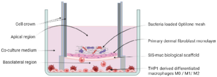
| Cells and culture medium | Fibroblasts from fascia biopsies cultivated in DMEM suppl. with FBS. THP-1 cells cultivated in THP-1 medium (RPMI-1640, L-glutamine, FBS, penicillin/streptomycin) and PMA. Differentiation into M0, M1 and M2 macrophages using specifically supplemented THP-1 medium |
| 3D cell culture technique | Collagen based scaffold derived from porcine intestine (SIS-muc) seeded with fibroblasts on the apical side. After 24 h, scaffolds were placed within cell crown inserts into medium (50:50 THP-1 medium and fibroblast medium) with differentiated macrophages (either M0, M1 or M2) and cultured for 11 days |
| Bacteria species | S. simulans and P. stutzeri mixed and cultivated for 48 h on polypropylene mesh (implant material) for biofilm formation |
| Bacterial challenge | Bacteria loaded polypropylene meshes placed on top of the fibroblasts Co-culture time 3 days |
| Sketch of the model |
 from Murkar et al. (2024) from Murkar et al. (2024)
|