
Figure 2:
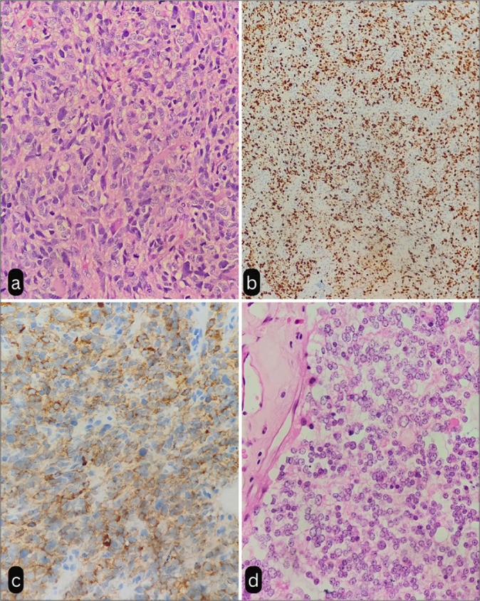
(a) Tumor composed of sheets of neoplastic tumor cells showing pleomorphism and increased mitosis suggestive of pineal parenchymal tumor of intermediate differentiation (H&E, ×40). (b) Tumor cells showing high Ki67 index (×10). (c) Tumor cells showing immunoreactivity to Synaptophysin (×40). (d) Photomicrograph showing uniform cells with round nuclei, moderate amount of eosinophilic cytoplasm, and absence of mitosis (H&E, ×40). H & E: Hematoxylin and Eosin Stain