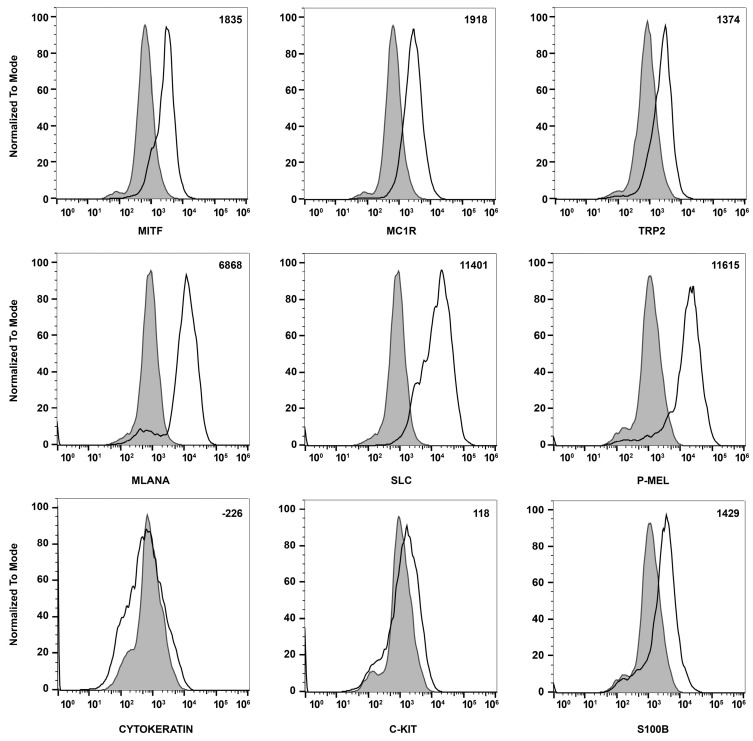
Figure 3.
Expression of melanocyte markers in ChMC. Mouse ChMC were examined for expression of Mitf, Mc1r, Trp2, Mlana, Slc, P-Mel, Pan Cytokeratin (RPE marker), c-Kit, and S100β using flow cytometry. The shaded areas show staining in the absence of primary antibody (secondary control), and the unshaded peaks show staining with primary antibody. The difference between the geometric means of primary antibody-stained cells and secondary antibody-stained control cells can be found in the top right corner of each graph. These experiments were performed at least 2 times with 3 different isolations of ChMC with similar results.

