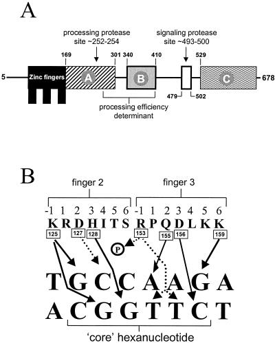
FIG. 2.
Schematic representation of PacC and its DNA binding specificity. (A) Functionally relevant regions in PacC are shown, with limits indicated by residue numbers. Interacting regions A, B, and C are required for maintaining the closed PacC conformation (41). The open box denotes the 24-residue signaling protease box (33). The approximate positions of the signaling protease and the processing protease cleavage sites are indicated by arrows. (B) Prediction of specific contacts between residues in the reading α-helix of PacC zinc fingers 2 and 3 and the PacC target hexanucleotide. Experimental evidence and modeling (43) strongly suggest the contacts indicated by the solid lines. Almost every base in both strands of the target site is predicted to establish specific contacts with PacC zinc finger residues. Dotted arrows indicate possible contacts of Asp127 and of Arg153, whose side chain can be modeled as contacting either the phosphate backbone or the O-4 atoms of both the T4′ and T5′ thymines. Note that finger 1 does not appear to be involved in specific contacts with DNA.

