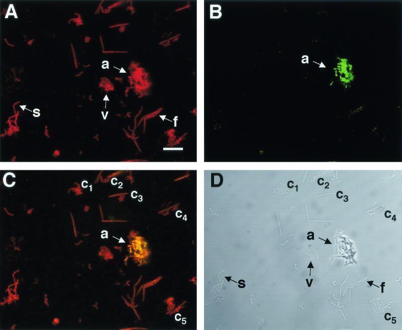
FIG. 3.
Four-genus mixture of oral bacteria stained with a specific probe by FISH and the nonspecific nucleic acid stain SYTO 59. (A) Confocal micrograph of a field of cells stained with the nucleic acid stain SYTO 59. The field of cells contains the following species: Actinomyces serovar WVA963 strain PK1259 (a), F. nucleatum PK1594 (f), S. gordonii DL1 (s), and Veillonella atypica PK1910 (v). (B) Confocal micrograph of the same field showing the location of the fluorescein isothiocyanate-labeled actinomyces-specific probe. The image demonstrates that the probe interacts only with actinomyces cells (a). (C) Overlay of confocal micrographs (A and B), demonstrating the specificity of the actinomyces probe. Areas of colocalization of fluorescein isothiocyanate and SYTO-59 markers appear yellow. The yellow actinomyces cells are in contact with other cells seen at the edges of the actinomyces cluster. Coaggregations (c1 to c5) of different species within the mixed culture are also visible. (D) Differential interference contrast image of the field of cells using transmitted light. The distinct morphologies of the various cells in the mixed culture are visible. Bar, 10 μm. Microscopic observations and image acquisition were performed on a TCS 4D system (Leica Lasertechnik GmbH, Heidelberg, Germany).

