
Figure 3.
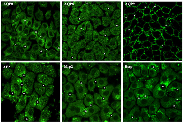
Confocal immunofluorescence for aquaporins and solute transporters in WIF-B cells. WIF-B cells were fixed, permeabilized, and labeled with anti-AQPs, AE2, Mrp2 or Bsep. Fluorescence localization was viewed by laser scanning confocal microscopy (see "Materials and Methods" for details). *, bile canaliculi structures.