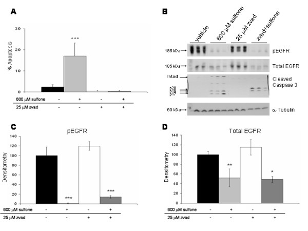
Figure 7.

Effect of the caspase inhibitor, ZVAD, on apoptosis and inhibition of EGFR. HT29 colon cancer cells were grown to confluence in medium containing 10% FBS followed by pretreatment with or without 25 μM zvad for 1 h. Cells were then treated with vehicle (0.2% DMSO) or 600 μM sulfone for 48 h. Cells were harvested and immunoblots were performed on cell lysates with antibodies raised against pEGFR (pY1068), total EGFR, and cleaved caspase 3; α-tubulin immunoblots of the same lysates served as loading controls. The graphs show morphological apoptosis results (A) 48 h Western immunoblot results (B) and densitometry of the pEGFR bands (C) and total EGFR bands (D). Data represent mean of triplicate samples ± SD; statistical significance is denoted by *p < 0.05, **p < 0.01 and ***p < 0.001. Results shown in figure are representative of 2 separate experiments each with triplicate samples.
