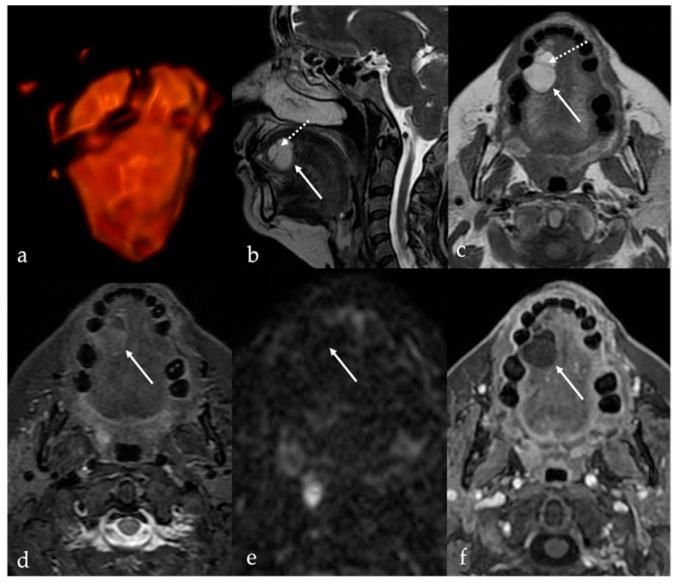
Figure 2.
Simple lipoma of the lingual body in a 59-year-old female patient. Magnetic resonance imaging (MRI) reveals an oval, well-defined mass (white arrows) located in the right side of the mobile tongue close to the right margin and the lingual apex. The mass (represented on direct volume rendering in (a)) shows intralesional fine septa (white dotted arrows) and high signal intensity (SI) on T2 sequence (b,c), homogenous intralesional signal saturation on Short Tau Inversion Recovery (STIR) sequence (d), lack of SI on diffusion-weighted imaging (e), and absence of enhancement after intravenous injection of gadolinium-based contrast medium (f), resembling subcutaneous adipose tissue.

