Figure 3.
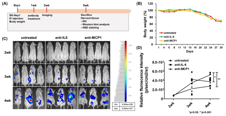
The suppression of tumor development and metastasis of HCC in mice by antibody treatment. NRG mice (5 weeks old/female) deficient in lymphocytes (T, B and NK cells) were used for in vivo experiments. The mice were injected intravenously through the tail with Luc-SK-Hep1 cells (5 × 10⁶ cells/mouse), divided into three groups, and treated after 1 week with intraperitoneal injections of DPBS, anti-IL-6 (2 μg/kg), or anti-MCP1 (1 μg/kg) twice a week until the end of this study. Tumors were visualized using an in vivo imaging system 2 weeks after the injection of the cancer cells. The overall scheme of the in vivo experiment (A), mouse body weight (B), the fluorescence intensity of luciferase in mice (C), and its quantitative graph (D) are shown. Data were analyzed by one-way ANOVA with Turkey’s multiple comparison test and significance between groups was indicated by a p-value of less than 0.05. * p < 0.05, ** p < 0.001.

