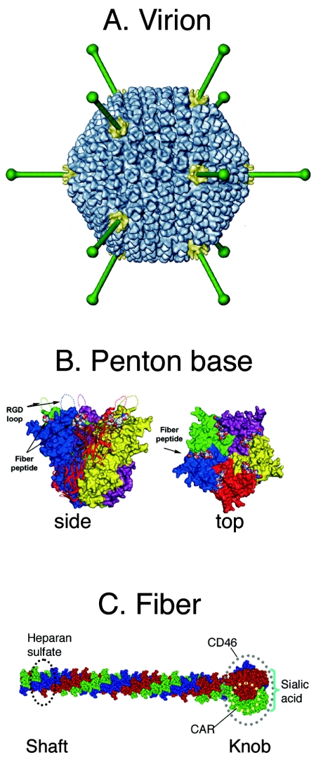
FIG. 1.
Adenovirus structure. (A) Virion structure at 17-Å resolution. Each of the 20 triangular faces of the capsid is composed of 12 copies of the hexon trimer (blue). At each fivefold vertex, a fiber (green) emerges from the pentameric penton base (yellow). The hexon capsid and penton base structures are derived from a cryoelectron microscopic image reconstruction of human adenovirus 5. Fibers are modeled from the atomic structure of the Ad2 fiber (95). Figure provided by Carmen San Martin. (B) Penton base. Side and top views, showing flexible RGD loops projecting from the surface. The insertion sites for fiber are also shown. (The figure is reprinted from reference 68 with permission of the publisher.) (C) Fiber structure and receptor binding sites. The fiber is a trimer whose monomers are indicated in red, blue, and green; the shaft is a tightly wound triple spiral; the knob is a more bulbous trefoil. The figure is modified from reference 95 with permission of the publisher.

