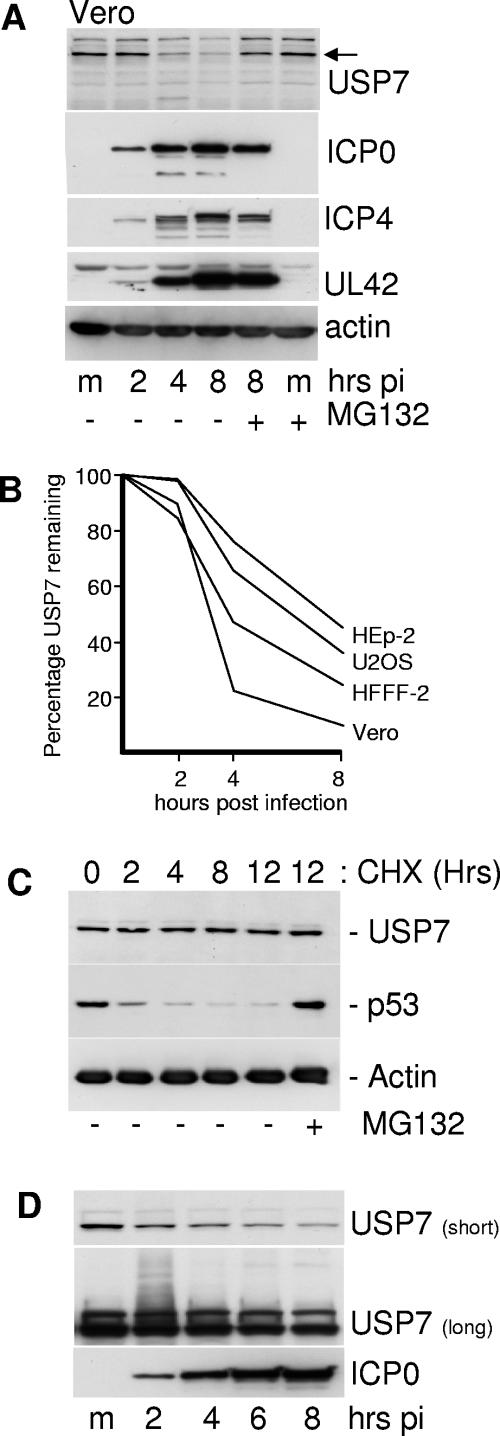
FIG. 3.
Levels of USP7 are reduced in a proteasome-dependent manner during HSV-1 infection of several cell types. (A) Vero cells were infected with wild-type HSV-1 (MOI of 20 PFU/cell) and samples were taken at 2, 4, and 8 h after virus adsorption. Whole-cell extracts were analyzed by Western blotting for USP7, immediate-early proteins ICP0 and ICP4, DNA replication protein UL42, and actin as indicated. m, mock-infected control. The two rightmost tracks contain samples from cells that had been treated with MG132 (10 μM final concentration) from the time of infection. (B) Vero, HFFF-2, Hep-2, and U2OS cells were infected with wild-type HSV-1 (MOI of 20 PFU/cell), and the levels of USP7 at 2, 4, and 8 h after infection were determined by Western blot analysis and densitometry as described in the text. (C) USP7 is stable in uninfected cells. HFFF-2 cells were treated with cycloheximide (100 μg/ml) and then samples were taken at the indicated time points and analyzed for USP7, p53, and actin. MG132 was present at 10 μM in the rightmost sample. (D) Detection of likely ubiquitinated forms of USP7 during HSV-1 infection. HFFF-2 cells were infected with wild-type HSV-1 (MOI of 20 PFU/cell) and then samples taken at 2-hour intervals were analyzed by Western blotting. The short exposure (10 seconds) demonstrates the decrease in USP7 levels that occurs during infection; the longer exposure (10 min) detects the smear of lower-mobility forms of USP7 in the 2-h sample.

