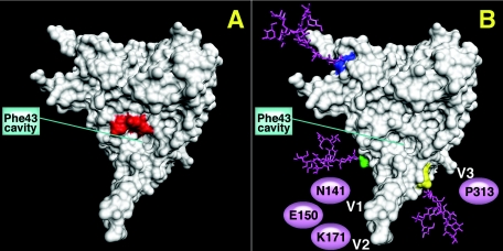
FIG. 1.
gp120 core structure with spatial location of the V1/V2/V3 loops representing mutant GDMR gp120 (A), where residues 473 to 476 (red) lining the CD4 Phe-43 binding cavity were mutated to alanine. (B) Mutant mCHO gp120 (CD4 binding face) with sugars (pink) attached on the core to residues H92 (dark blue), Q114 (green), and I423 (yellow). The putative location of V1 and V2 loops with three sugars at position N141, E150, and K171 and the putative location of the V3 loop with one sugar at position P313 are also represented. The monomer structure is from the Protein Data Bank of HXBc2 gp120 by Kwong et al. (24). The images were made using the PMV program by C. Corbaci. R. Cardoso attached the coordinates for the sugars at residues H92, Q114, and I423 on the gp120 core representing the extra glycans on the core of mCHO gp120 mutant.

