Abstract
Using PCR mutagenesis to disrupt the NXT/S N-linked glycosylation motif of the Env protein, we created 27 mutants lacking 1 to 5 of 14 N-linked glycosylation sites within regions of gp120 lying outside of variable loops 1 to 4 within simian immunodeficiency virus strain 239 (SIV239). Of 18 mutants missing N-linked glycosylation sites predicted to lie within 10 Å of CD4 contact sites, the infectivity of 12 was sufficient to measure sensitivity to neutralization by soluble CD4 (sCD4), pooled immune sera from SIV239-infected rhesus macaques, and monoclonal antibodies known to neutralize certain derivatives of SIV239. Three of these 12 mutants (g3, lacking the 3rd glycan at position 79; g11, lacking the 11th glycan at position 212; and g3,11, lacking both the 3rd and 11th glycans) were approximately five times more sensitive to neutralization by sCD4 than wild-type (WT) SIV239. However, these same mutants were no more sensitive to neutralization than WT by pooled immune sera. The other 9 of 12 replication-competent mutants in this group were no more sensitive to neutralization than the WT by any of the neutralizing reagents. Six of the nine mutants that did not replicate appreciably had three or more glycosylation sites eliminated; the other three replication-deficient strains involved mutation of site 15. Our results suggest that elimination of glycan attachment sites 3 and 11 enhanced the exposure of contact residues for CD4. Thus, glycans at positions 3 and 11 of SIV239 gp120 may be particularly important for shielding the CD4-binding site from antibody recognition.
Vaccine-induced protection against a number of viral pathogens correlates well with neutralizing antibody (Ab) titers (2, 17, 20, 53). Some have suggested that the most effective vaccine against human immunodeficiency virus (HIV) is likely to be one capable of eliciting strong, broadly reactive neutralizing antibody responses to Env as well as broad-spectrum cellular immune responses. The poor immunogenicity of the Env spike, however, is a major obstacle to the engineering of such a vaccine. One of the features of Env that contributes to its ability to escape immune recognition is its high level of glycosylation. HIV type 1 (HIV-1) gp120 typically contains more than 20 N-linked glycosylation sites (15) and 8 O-linked sites (3). A survey performed by Myers and Lenroot of approximately 10,000 protein sequences in the SWISS-PROT library with at least one potential N-linked glycosylation site found that the number of glycosylation sites in HIV-1RF ranked in the top 10 proteins with respect to the degree of carbohydrate modification (41).
N- and O-linked glycosylation of the Env precursor protein gp160 occurs during translation. Oligomerization into trimers also occurs while the nascent protein transiently resides within the endoplasmic reticulum. The newly added carbohydrate moieties are further trimmed prior to the transport of the Env oligomer to the Golgi network, where subsequent glycan diversification occurs. Formation of the viral spike is completed within the Golgi network, where gp160 is cleaved into the surface protein gp120 and the transmembrane fusion protein gp41. These two noncovalently linked subunits form heterotrimers, which become incorporated into the viral membrane upon budding.
Certain glycosylation sites on cellular or viral proteins are critical to proper protein folding, intracellular stabilization, and protection against cellular proteases (10, 13, 14, 43, 46). Loss of particular glycans can also affect viral infectivity, possibly through structural alterations that influence the ability of the glycoprotein to bind its receptors, monomer interactions within the trimer, or interactions of the surface and transmembrane fusion proteins (31, 42, 55). However, many single and multiple glycosylation sites have been shown to be dispensable to viral replication within HIV-1 gp41 (11, 23) and gp120 of both HIV-1 (4, 16, 25, 26, 34, 51) and simian immunodeficiency virus (SIV) (39, 47, 48). The dispensability of some of these N-linked glycans to viral replication and the greater sensitivity of some mutants missing glycan attachment sites to antibody-mediated neutralization (4, 16, 27, 34, 36, 45, 48, 51) suggest that these glycans may also serve to shield the spike from recognition by antibodies.
Variations in the number or location of glycosylation sites, particularly within the V1/V2 and V3 loops but also on the “silent face” of gp120, often correlate with altered sensitivity to neutralizing antibodies (1, 7, 18, 35, 44, 50, 54). Just as the acquisition of particular N-linked sites decreases neutralization sensitivity, elimination of N-linked sites at the same or nearby locations has been shown to increase neutralization sensitivity within both HIV-1 and SIV strains, particularly in the V1/V2 and V3 loops (4, 16, 27, 34, 45, 48, 51). Less is known with regard to the effects of mutagenesis of glycosylation sites outside of the V1/V2 and V3 loops on neutralization sensitivity of SIV or HIV.
To address the possibility that elimination of N-linked glycans located within the better-conserved core of gp120 might expose relatively conserved domains, we created 27 mutant strains of SIV strain 239 (SIV239) that lack one or more of 14 core N-linked glycosylation sites. The mutants that lacked N-linked glycans proximal to receptor binding sites were of primary interest. We chose to work with SIV239 in order to allow future studies relating to immunopathogenesis upon experimental rhesus macaque infection. Our studies demonstrate that elimination of single glycosylation sites within the core are in general well tolerated. Based upon soluble CD4 (sCD4) neutralization sensitivity, we have identified two glycosylation sites that may limit access to CD4 and thus play a role in shielding the CD4-binding site (CD4bs) from antibody recognition.
MATERIALS AND METHODS
Viral genome plasmids.
Mutations in env were introduced into proviral SIV239 DNA using pSP72-239-3′, a plasmid containing the 3′ half of SIV239. The 3′ half of SIV239 contains the viral env gene, the open reading frames for tat, rev, nef, vpx, vpr, and vpu, and the 3′ long terminal repeat. The 5′ half of SIV239 contains the 5′ long terminal repeat and the gag, pol, and vif genes. The two halves of SIV239 were ligated together at the SphI unique restriction site to obtain full-length virus.
Site-specific mutagenesis.
The NXT/S-to-QXT/S mutations in env were made using a QuickChange site-directed mutagenesis kit (Stratagene, La Jolla, CA). To avoid PCR-induced changes outside of env, the Bsm1 fragment (1,710 bp) containing the mutation was inserted back into pSP72-239-3′ and sequenced using an ABI377 automated DNA sequencer by using dye terminator cycle-sequencing chemistry as specified by the manufacturer (Perkin-Elmer Inc., Foster City, CA).
Virus stocks and cell culture.
For the generation of viral stocks, 10 μg of the 5′ and 3′ clones of SIV239 were digested with SphI. Each 3′ clone was ligated with the 5′ clone using T4 DNA ligase. The ligated DNA was used to transfect 293T cells by the calcium phosphate method (Promega Biotech, Madison, WI). Cell culture supernatant was harvested on day 3 posttransfection, and viral content was quantitated by p27 using a commercial antigen capture assay (Coulter Corp., Hialeah, FL). 293T and CEMx174 cells were maintained as previously described (38, 40).
Viral growth curves.
To obtain growth curves in CEMx174 cells, viral stocks containing 10 ng of p27 were added to 5 × 10⁶ cells at a concentration of 2.5 × 10⁶ cell/ml and incubated at 37°C and 5% CO2 for 1 h. The cells were then pelleted and resuspended in 5 ml of fresh RPMI 1640-10 medium. Five milliliters of the supernatant was replaced with fresh medium every 3 to 4 days. The cell-free supernatant was harvested on the days indicated, and the amount of p27 was quantitated.
Infectivity assays.
Each viral strain was serially diluted 1:2 and mixed with 5 × 10⁴ indicator cells (CEMx174 stably transfected with a tat-responsive, secreted alkaline phosphatase [SEAP] reporter gene) (37). Supernatants from each well were collected 48 to 72 h after infection, and SEAP activity was measured by using a PhosphaLight kit (Applied Biosystems, Foster City, CA).
Immunoblotting.
For immunoblots of virion-associated Env and Gag proteins, supernatants of transfected 293T cells were collected at day 3 posttransfection. The entire volume of supernatant was concentrated using Amicon ultracentifugal filter devices (Millipore, Bedford, MA) to obtain approximately 1 ml of concentrated virus. One milliliter of concentrated supernatant was centrifuged at high speed for 2 h. The supernatant was removed, and the viral pellet was washed once with phosphate-buffered saline and then resuspended in 50 μl of lysis buffer (50 mM Tris-HCl, pH 8.0, 150 mM NaCl, and 1% NP-40 and one Complete tablet [a protease inhibitor cocktail]) (Roche Diagnostics, Mannheim, Germany). Viral proteins containing 50 ng of p27 were separated by sodium dodecyl sulfate-polyacrylamide gel electrophoresis followed by transfer to a polyvinylidene difluoride membrane (Millipore, Bedford, MA). Due to the high band intensity of p27, the blot was first incubated with a rabbit polyclonal antiserum to SIV251 gp120 (Microbix Biosystems Inc., Toronto, Ontario, Canada) at a concentration of 1:1,000 followed by a horseradish peroxidase-conjugated secondary anti-rabbit antibody and developed by chemiluminescence using a PhosphorImager. The blot was then blocked with 5% milk in phosphate-buffered saline, pH 7.4, and 0.05% Tween 20 and then incubated with a murine monoclonal antibody (MAb) to p27, 2F12 (National Institutes of Health AIDS Research and Reference Reagent Program), followed by a horseradish peroxidase-labeled goat-anti mouse antibody (Santa Cruz Biotech., Santa Cruz, CA), and bands were detected by chemiluminescence using a PhosphorImager. In some experiments, blots were incubated with anti-gp41 MAb KK41 (24) to detect gp41 and/or unprocessed gp160; these same blots were then sequentially stripped and reprobed with polyclonal anti-gp120 Ab and anti-p27 Ab 2F12.
Neutralization assay.
The neutralization sensitivity of each mutant was tested by using the SEAP reporter cell assay as described previously (37). Aliquots of virus stocks used in these experiments were subject to serial twofold dilutions, and their titers were determined on SEAP reporter cells. The virus equivalent to 2 ng of p27 capsid protein was chosen as the lowest level of input sufficient to give a clear SEAP signal within the linear range for most of the virus strains. Those strains with infectivity in the 20 to 30% range required a higher input, usually 3 to 5 ng/well. SEAP activity was measured at 72 h for viruses that possessed levels of infectivity close to that of wild-type (WT) virus, and others with low infectivity required longer incubations (up to 7 days for some). The percent neutralization of SIV239 did not vary significantly over multiple days postinfection. The neutralization assay was set up as described previously (22). Neutralization activity for sCD4, sera, and MAbs was measured in duplicate and reported as the average of two measurements, with error bars representing the standard deviations. The percent SEAP was calculated as the amount of SEAP produced in the presence of either sCD4, immune serum, or MAb divided by the amount of SEAP produced in the absence of any inhibitor.
RESULTS
Location of N-linked glycosylation sites selected for mutagenesis.
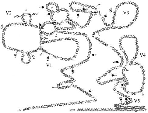
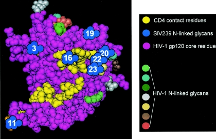
We aligned the amino acid sequence of the SIV239 gp120 core with that of HIV-1HXBc2, the gp120 molecule from which the published crystal structure was derived (30). Alignment of SIV239 with HIV-1 was facilitated by the previously published alignment of the gp120 core of HIV-1HXBc2 with that of HIV-2ROD, a strain with substantial sequence homology to SIV239 (30). Using Cn3D 4.1 (http://www.ncbi.nlm.nih.gov/Structure/CN3D/cn3d.shtml), we were able to locate amino acids in HIV-1HXBc2 that aligned with SIV239 gp120 amino acids within the crystal structure of gp120 (29). With this method, we identified 14 potential N-linked glycosylation sites within the gp120 core structure and mapped their presumed location onto the crystal structure for HIV-1. The locations of these 14 N residues within SIV239 gp120 are shown in Fig. 1. We selected N-linked sites in SIV239 gp120 that appeared to lie within 10 Å of residues thought to make contact with CD4. We hypothesized that carbohydrates at these sites may help shield the CD4-binding domain from antibody recognition. The locations of seven potential N-linked sites that are predicted to lie within 10 Å of CD4 contact residues are shown in Fig. 2. Each N-linked glycosylation site was mutagenized using PCR to change the N of the NXT/S sequence for N-linked glycosylation to a structurally similar Q. All N-to-Q substitutions were engineered such that a minimum of two nucleotide changes would be required for the sequence to revert to an N (47).
FIG. 1.
Schematic of SIV239 gp120. Amino acids representing the entire primary sequence of SIV239 gp120 were drawn schematically based on the known disulfide bonds for HIV-1 (33) and the hypothetical disulfide bonds for SIV and HIV-2 (21). The variable regions were based upon the designations defined previously by Choi et al. (8), and the V3 region corresponds to the analogous V3 loop of HIV-1. All potential N-linked glycosylation sites are indicated and numbered g1 through g23. The blackened symbols for glycosylation indicate the 14 that were eliminated singly or in various combinations in our mutant strains.
FIG. 2.
Deduced locations of N-linked glycosylation sites of SIV239 gp120 which appear to lie proximal to CD4 contact residues. The N-linked sites are superimposed upon the crystal structure of HIV-1HXBc2 (30) and are designated by a numbered blue cluster indicating the number of the glycosylation site in SIV239 gp120. These sites were located using program Cn3D 4.1. The variously colored clusters in the figure represent HIV-1HXBc2 N-linked glycosylation sites. The yellow highlighted residues are those of HIV-1HXBc2 that make contact with CD4.
Infectivity, replication in culture, and neutralization of mutant strains with N-linked glycosylation sites eliminated that appear to lie within 10 Å of CD4 contact residues.
N-linked sites predicted to be proximal to CD4 contact sites were eliminated in single and multiple combinations to create 18 mutant strains of SIV239 (Table 1). Infectivity was measured in an assay designed to estimate a single cycle of viral infection (37). The seven mutants that were missing only a single N-linked site were all infectious and replicated well in cell culture.
TABLE 1.
Infectivity, in vitro replicative capacity, and IC50 values of sera and sCD4 against mutant strains lacking glycosylation sites proximal to CD4 contact residuesa
| Viral strain | Infectivity (% of WT) | Growth in tissue culture | IC50 sera (inverse dilution) | IC50 sCD4 (ng/μl) |
|---|---|---|---|---|
| SIV239 | NA | Yes | <20 | 17.8 |
| g3 | 170 | Yes | <20 | 4.0 |
| g11 | 8 | Yes | <20 | 3.6 |
| g16 | 35 | Yes | 15 | 39.0 |
| g19 | 65 | Yes | 20 | 6.0 |
| g20 | 33 | Yes | <20 | 24.0 |
| g22 | 69 | Yes | <20 | 16.0 |
| g23 | 82 | Yes | <20 | 12.0 |
| g3,11 | 14 | Yes | <20 | 3.5 |
| g19,20 | 12 | Yes | <20 | 22.0 |
| g22,23 | 59 | Yes | <20 | 9.0 |
| g14,22,23 | 2 | Yesb | NA | NA |
| g16,22,23 | 0 | No | NA | NA |
| g19,20,22,23 | 33 | Yes | 30 | 32 |
| g3,11,19,20 | 17 | Yes | >20 | 9.4 |
| g16,19,20,22,23 | 0 | No | NA | NA |
| g14,19,20,22,23 | 0 | No | NA | NA |
| g15,19,20,22,23 | 0 | No | NA | NA |
| g11,19,20,22,23 | 2 | No | NA | NA |
NA, nonapplicable due to low to no infectivity.
Replication was noted by day 32, and sequencing of reverse transcription-PCR products derived from cell-free virus revealed nonsynonymous substitutions that were likely compensatory. The mutants in boldface type are those with enhanced neutralization sensitivity to sCD4.
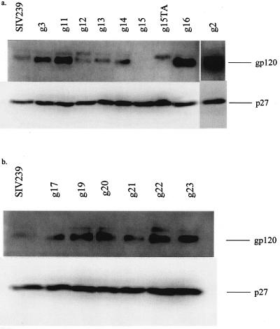
On immunoblotting analysis of viral protein, a band representing gp120 derived from each of these mutants migrated slightly lower than WT SIV239 gp120, indicating the utilization of each of these sites in our cell culture system (Fig. 3a and b). Some of the mutants appear to have differing Env content compared to that of the WT.
FIG. 3.
(a and b) Virion-associated Env expression of WT SIV239 and mutant virions missing one N-linked glycosylation site produced in 293T cells. (a) Immunoblot of WT SIV239 virion protein and mutants g2, g3, g11, g12, g13, g14, g15, g15TA, g16, and g2. (b) WT SIV239 and mutants g17, g19, g20, g21, g22, and g23. 293T cells were transiently transfected with proviral DNA of WT SIV239 and each of the glycosylation mutants of SIV239. Lysed viral pellets from these transfections were generated, and 50 ng of p27 from each was loaded per lane. gp120 and p27 were detected by SIV anti-gp120 polyclonal antibody (top panels in a and b) and 2F12 (bottom panels in a and b), respectively. Detection of gp120 and p27 was done sequentially: the membrane was first probed with antisera to SIV gp120, and results were visualized using chemiluminescence and then blocked again and probed with 2F12. This was to avoid quenching of the signal for gp120 by the more intense signal obtained with 2F12. Mutant g2 was cut from the blot, and its chemiluminescent signal was detected separately due to the intensity of the gp120 band.
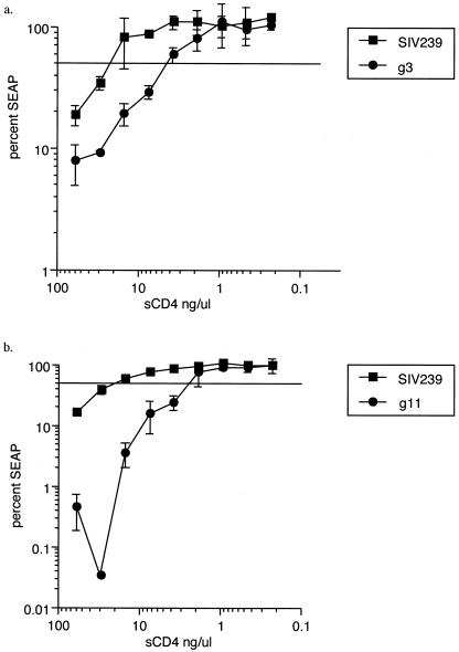
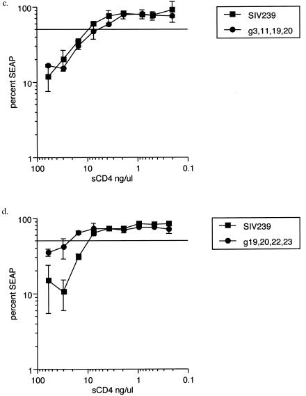
Three of 12 replication-competent mutants, g3, g11, and g3,11, displayed a fivefold increase in sensitivity to neutralization by sCD4 (Table 1 and Fig. 4a and b). The remaining nine replication-competent mutants in this group were no more sensitive to neutralization by sCD4 than the WT, including two mutants missing four N-linked sites: g19,20,22,23 and g3,11,19,20 (Fig. 4c and d). All 12 replication-competent mutants were no better neutralized using pooled immune sera obtained from SIV239-infected rhesus macaques than WT SIV239.
FIG. 4.
Neutralization of four mutants of SIV239 and SIV239 WT. (a) Neutralization of the WT and g3 by sCD4; (b) neutralization of the WT and g11 by sCD4. (c) Neutralization of WT and g3,11,19,20 by sCD4. (d) Neutralization of WT and g19,20,22,23 by sCD4. Percent SEAP is the amount of SEAP produced in the presence of a particular concentration of sCD4 relative to the amount of SEAP produced in the absence of sCD4 or sera. A line is drawn through the 50% SEAP point on the y axis to indicate that the corresponding x value is the 50% inhibitory concentration (IC50) for each virus.
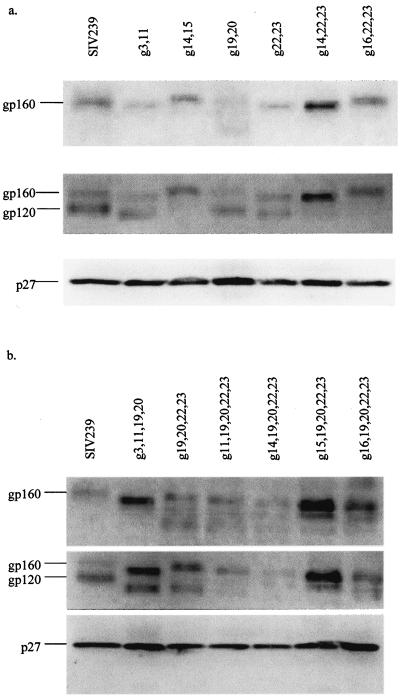
Many of the mutants with three or more N-linked sites eliminated in particular combinations were not infectious and were unable to grow in cell culture. That the loss of these glycans resulted in aberrant Env protein is supported by the unusual pattern of virion-associated Env observed in immunoblots (Fig. 5a and b). All of these mutants displayed a slower migrating band that reacted with a monoclonal antibody for gp41, suggesting that these mutants incorporated unprocessed Env and little or no mature gp120.
FIG. 5.
Virion-associated Env expression of WT SIV239 and mutant virions missing combinations of N-linked glycosylation sites produced in 293T cells. Viral pellets were obtained as described in the legend of Fig. 3, and 50 ng of p27 was loaded per lane. (a) Mutants lacking two or three sites. The top panel was immunoblotted with KK41, a MAb to SIV gp41, the middle panel was immunoblotted with polyclonal antibody to SIV gp120, and the bottom panel was immunoblotted with 2F12, a MAb to p27. (b) Mutants lacking four or five N-linked sites. The top, middle, and bottom panels are arranged in the same manner as described above in panel a.
Neutralization of two mutants using a panel of MAbs to SIV239 gp120.
To further explore whether the elimination of the glycans in these mutants resulted in enhanced sensitivity to neutralization, we used a panel of rhesus macaque-derived MAbs directed to SIVgp120. These antibodies were kindly provided by both James Robinson and James Hoxie. Competition analysis with antibodies of known specificity was used to map the specificities of these 10 MAbs (9); some, presumed to be conformational, were attributed to a particular competition group rather than a specific region of gp120. This panel contains MAbs that map to both discontinuous and linear epitopes that span much of gp120 and that have been shown to neutralize certain neutralization-sensitive derivatives of SIV239 such as SIV316 (9, 12, 22). None of the MAbs in this panel have been characterized as CD4bs specific. Neither g3,11 (which was approximately fivefold more sensitive to sCD4 than WT virus) nor g19,20,22,23 (Fig. 2) was more sensitive than WT SIV239 to neutralization by any of the MAbs (Table 2).
TABLE 2.
Neutralization of two glycosylation mutants using a panel of 10 MAbs capable of neutralizing SIV239-derived strainsa
| Viral strain | MAb IC50 titer (ng/μl)
|
|||||||||
|---|---|---|---|---|---|---|---|---|---|---|
| 1.9C | 1.10A | 3.4E | 3.10A | 3.11H | 5.5B | 22A | 59C | E31 | C26 | |
| SIV239 | >14.6 | >62.5 | >26.9 | >39.8 | >80 | >16 | >Neat | >Neat | >41.8 | >50 |
| g3,11 | >14.6 | >62.5 | >26.9 | >39.8 | >80 | >16 | >Neat | >Neat | >41.8 | >50 |
| g19,20,22,23 | >14.6 | >62.5 | >26.9 | >39.8 | >80 | >16 | >Neat | >Neat | >41.8 | >50 |
| SIV316 | 0.001 | 0.004 | 0.002 | 0.1 | 0.005 | 0.04 | ND | ND | 0.0002 | 0.0008 |
See references 9 and 12. The titers are IC50 for each antibody against WT SIV239, g3,11, and g19,20,22,23. The MAbs have the following specificities based upon competition studies (9): 1.10A, V4; C26, V4; E31, competition group VI; 1.9C, competition group VI; 3.4E, competition group V; 3.11H, V3; 5.5B, V2; 3.10A, V1; 59C2, V2 (positions 171 to 190); and 22A, V1 (positions 111 to 130). The first eight MAbs were purified with known concentrations generously provided by James Robinson. The last two MAbs (59C2 and 22A) were hybridoma supernatants generously provided by James Hoxie. The 50% neutralization values for SIV316 are derived from the work of Johnson et al (22). ND, not done.
Infectivity, replication in culture, and neutralization of mutant strains with N-linked glycosylation sites eliminated that appear to lie far from CD4 contact residues.
We analyzed the infectivity, replication kinetics, and neutralization profiles of nine glycosylation mutants of SIV239 in which the eliminated N-linked sites were predicted to lie greater than 10 Å from CD4 contact residues (Table 3). Individual elimination of all but one of the glycans was well tolerated by the virus. We were unable to detect infectivity in one mutant, g15, and this mutant strain did not replicate in long-term culture. To test whether loss of N-linked glycan or simply loss of asparagine at that site was responsible, we made a second strain in which glycosylation of site 15 was eliminated by substituting an alanine for threonine (NXT to NXA); this mutant was named g15TA to distinguish it from g15 (QXT). Mutation of the N-linked glycosylation site by changing the T of the NXT sequence to an A resulted in the same replication-defective phenotype. A second strain, with mutations at glycosylation sites 14 and 15 (g14,15), was also noninfectious and unable to replicate in long-term culture. On immunoblotting, gp120 of the six replication-competent mutant strains migrated discernibly lower than WT SIV239, indicating that these N-linked sites were utilized in our cell culture system (Fig. 3a and b). Mutant g15 had no detectable viral pellet-associated Env protein by immunoblotting, whereas g15TA did have a higher-molecular-weight species of Env, migrating at 160 kDa (Fig. 3a). Neutralization sensitivity to sCD4 or pooled rhesus SIV239 immune sera was not significantly enhanced compared to wild-type SIV239 sensitivity for any of the six infectious mutants in this group (Table 3).
TABLE 3.
Infectivity, in vitro replicative capacity, and IC50 values of sera and sCD4 against mutant strains lacking glycosylation sites distal to receptor contact residuesa
| Viral strain | Infectivity (% of WT) | Growth in tissue culture | IC50 sera (inverse dilution) | IC50 sCD4 (ng/μl) |
|---|---|---|---|---|
| SIV239 | NA | Yes | <20 | 17.8 |
| g2 | 28 | Yes | <20 | 7.5 |
| g12 | 46 | Yes | 20 | 6.3 |
| g13 | 170 | Yes | 20 | 5.0 |
| g14 | 5 | Yes | 23 | 14 |
| g15 | 0 | No | NA | NA |
| g15TA | 0 | No | NA | NA |
| g17 | 41 | Yes | 20 | 11.0 |
| g21 | 120 | Yes | 12 | 11.0 |
| g14,15 | 0 | No | NA | NA |
NA, nonapplicable. Mutants were tested for their ability to grow in long-term tissue culture in the CEMx174 cell line.
Mutants lacking glycosylation sites lying proximal to the second receptor binding site.
Glycosylation sites at the 11th, 21st, 22nd, and 23rd N-linked attachment sites in SIV239 gp120 are predicted to lie proximal to residues K121, R419, K421, and Q422 identified in HIV-1 for second-receptor binding (30). We evaluated the neutralization sensitivity of mutants g11, g21, g22, and g23 against three monoclonal antibodies that block CCR5 binding of CD4-independent strains of SIV (12). None of these mutants displayed increased sensitivity to neutralization with these antibodies relative to parental SIV239 (Table 4).
TABLE 4.
IC50s of MAbs 7D3, 8C7, and 4E11a
| Viral strain | Inverse dilution MAb IC50 (ng/μl)
|
||
|---|---|---|---|
| 7D3 | 8C7 | 4E11 | |
| SIV239 | >20 | >20 | >20 |
| g11 | >20 | >20 | >20 |
| g21 | >20 | >20 | >20 |
| g22 | >20 | >20 | >20 |
| g23 | >20 | >20 | >20 |
IC50s of MAbs 7D3, 8C7, and 4E11 (hybridoma supernatants) which block SIV316 gp120:CCR5 binding (12) against mutant strains lacking glycosylation sites proximal to the CCR5 binding site.
DISCUSSION
A number of previous studies have shown that elimination of particular glycosylation sites in gp120 in HIV-1 and SIV results in enhanced sensitivity to neutralization by a wide variety of antibodies and other reagents. However, most of these studies have focused on glycosylation variation within variable regions such as V1/V2 and V3 (1, 7, 19, 35, 44, 50, 54). In this study, we have created a number of mutants of SIV239 missing N-linked glycans, either singly or in particular combinations, that lie within the conserved core of gp120 and evaluated the contribution of each of these glycans to neutralization resistance. Seven sites were predicted to lie proximal to CD4 contact residues and therefore could conceivably limit access of antibodies and possibly CD4 itself. Analysis of 27 mutants revealed that 3 mutants (g3, g11, and g3,11) were five times more sensitive to neutralization by sCD4 than their parental strain, SIV239. These three mutants were no more sensitive to immune sera or a panel of 10 monoclonal antibodies than WT virus.
A crystal structure of unliganded, fully glycosylated SIV gp120 has been recently elucidated (5, 6). Of the seven SIV239 gp120 glycans that we presumed were proximal to CD4 contact sites relative to the CD4-binding loop of unliganded SIV gp120 (5, 6), only glycan 3 would be positioned in such a way in the structure as to sterically interfere with CD4 binding (B. Chen and S. Harrison, personal communication). Glycan 11, positioned at the stem of the V1/V2 loop, is not in a location that would be expected to block CD4 binding but is likely to impact upon movement of the V1/V2 loop (26). Glycans 16 and 19 appear to be oriented away from the initial area of CD4 contact and lie distal to it. Glycans 20, 22, and 23 form a sugar cluster designated cluster III in the unbound SIV gp120 structure (5). This cluster is also in a region of the outer domain that would not be expected to interfere with CD4 binding. Comparison of the crystal structures of unliganded SIV gp120 with the CD4-bound HIV-1 gp120 structure suggests that binding of CD4 to the CD4-binding loop may cause a large sequential reorganization of regions of the inner domain (5, 6). These rearrangements therefore presumably result in relocation of some or all of these glycans to regions in closer proximity to CD4 contact sites.
Glycosylation site 3, at the amino-terminal end of SIV239 Env (position 79), is found in 0% of HIV-1/SIVcpz and 25% of HIV-1/SIVmm sequences in the Los Alamos Database (32). Removal of this glycosylation site not only increases sensitivity of SIV239 to neutralization by sCD4 but also increases infectivity approximately twofold. It is positioned close to the region into which CD4 must enter to make contact with the CD4-binding loop in the unliganded SIV gp120 core structure (B. Chen and S. Harrison, personal communication), supporting the possibility that it creates some measure of steric inhibition to CD4 binding.
Glycosylation site 11, located at the stem of the V1/V2 loop, aligns with glycan 197 in the CD4-independent ADA isolate described previously by Kolchinsky et al. (27). That group showed that this HIV-1 isolate, solely on the basis of elimination of this one glycosylation site, was globally sensitive to neutralization with CD4-induced antibodies, CD4bs antibodies, and antibodies to the V3 loop as well as to gp41. Given the location of this glycan, those authors concluded that its elimination resulted in a change in gp120 conformation that allowed access to antibodies recognizing a wide variety of regions. Our SIV239 mutant, g11, did not demonstrate enhanced neutralization sensitivity to immune sera, suggesting that a conformational change leading to exposure of multiple sites on gp120 was unlikely in this mutant. However, the lowered infectivity (8%) of this mutant compared to that of WT virus supports the possibility of an alteration in gp120 conformation within the context of the trimer. Loss of this glycan might allow more favorable thermodynamics of binding for sCD4 and/or better exposure of CD4-binding regions. The degree of neutralization sensitivity to various gp120 antibodies and ligands in strains lacking this V1/V2 stem glycan is likely to be context dependent as it appears to be in strains of HIV-1 (34, 52).
Mutant g3,11 demonstrates neutralization sensitivity and infectivity (14%) similar to those of g11. It is also resistant to neutralization by a panel of MAbs that recognize a number of conformational epitopes in various regions of the spike as well as linear epitopes in variable loops V1 through V4. We do not know if our panel contains MAbs that recognize the CD4-binding site. It is possible that increased exposure of residues of the CD4-binding site resulting from the g3,11 mutation could result in increased susceptibility to neutralization by CD4bs antibodies, but such Abs may not have been present in the panel. Further testing of this possibility will require isolation of SIV-specific anti-CD4bs MAbs.
The lack of sensitivity of these three mutants to neutralization by immune sera from infected monkeys may be explained by the absence of potent CD4bs antibodies in monkeys infected with SIV239. It is likely that little if any potently neutralizing CD4bs antibodies are raised during natural SIV infection due to an energetic barrier contributed by the size of the antibody footprint as well as steric constraints on antibody binding imposed by glycosylation and oligomeric occlusion (28).
Removal of the 3rd and 11th glycan enhanced neutralization sensitivity to sCD4, yet removal of two additional glycans (to create the g3,11,19,20 strain) resulted in a loss of this sensitivity. This raises the possibility that the gp120 structure resulting from these additional changes is altered in such a way that the binding of CD4 is hampered. Structural alteration is suggested both by decreased infectivity (17% of that of WT virus) and prominence of a higher-molecular-weight band migrating at 160 kDa on Western blots of virion protein.
Edinger et al. derived MAbs that blocked recognition of CCR5 (12). These antibodies were raised in mice by immunization with whole cells infected with a CD4-independent strain of SIV. Their binding specificity was determined by their ability to block binding of a strain of CD4-independent SIV to rhesus CCR5-expressing 293T cells. These antibodies were able to neutralize CD4-independent SIV strains and did not compete with anti-V3 loop antibodies, suggesting that they may interact directly with a conserved chemokine receptor region in SIV. Mutational analysis of HIV-1 has demonstrated that residues Lys121, Arg419, Lys421, and Gln422 are important for CCR5 interaction (49). To locate glycosylation sites proximal to these residues, we used the crystal structure of HIV-1HXBc2. SIV239 glycosylation sites 11, 21, 22, and 23 were presumed to lie proximal to the CCR5 binding site using this analysis. However, mutants g11, g21, g22, and g23 on the neutralization-resistant SIV239 continued to be resistant to neutralization with three of the CCR5-blocking antibodies described previously (12). Removal of only one of these glycans is unlikely to create the structural changes that are thought to be present in CD4-independent strains of SIV and HIV that allow access to regions of second-receptor binding in the absence of CD4 (12, 26).
Interestingly, the majority of mutants with single gp120 core glycosylation site knockouts were replication competent. The only exception to this was glycosylation site 15. Loss of this single glycan resulted in complete loss of infectivity. Substitution of the T of the NXT glycosylation sequence to create an alternate glycosylation mutant, g15TA, also resulted in loss of infectivity. Mutant g15 had no detectable viral pellet-associated Env protein by immunoblotting; mutant g15TA did have a higher-molecular-weight band migrating at 160 kDa. These findings suggest that the lack of glycosylation at this site creates a lethal defect in Env processing and incorporation, but the N at position 282 may also contribute to Env stability and/or incorporation independent of its role as a glycan attachment moiety. Our findings are consistent with those of Ohgimoto et al., who created mutants identical to g15 and g15TA that were also noninfectious (42).
In summary, we have created a number of infectious mutants of SIV239 missing N-linked glycosylation sites within the core of gp120 and evaluated the contribution of these glycans to neutralization sensitivity. We have demonstrated a fivefold increase in sensitivity to sCD4 in three mutants of SIV239 that lack the 3rd and 11th glycans, which are thought to lie proximal to the CD4-binding site. Although we were not able to show increased neutralization by any of the antibodies in our panels, it is still reasonable to think that these glycans help to shield the CD4-binding site from antibody recognition; decreased affinity for CD4 would then be the price that the virus has to pay for limiting antibody accessibility.
Acknowledgments
We thank James Robinson and James Hoxie for providing MAbs to SIV Env and Bing Chen for discussions of the locations of N-linked glycosylation sites within his crystal structure of unliganded SIV gp120.
This work was supported by Public Service grants AI51158 (C.P.) and AI25328 (R.C.D.) and by base grant support to the New England Primate Research Center (RR00168).
REFERENCES
- 1.Back, N. K. T., L. Smit, J. DeJong, W. Keulen, M. Schutten, J. Goudsmit, and M. Tersmette. 1994. An N-glycan within the human immunodeficiency virus type 1 gp120 V3 loop affects virus neutralization. Virology 199:431-438. [DOI] [PubMed] [Google Scholar]
- 2.Bancroft, W. H., R. M. Scott, K. H. Eckels, C. H. J. Hoke, T. E. Simms, K. D. Jesrani, P. L. Summers, D. R. Dubois, D. Tsoulos, and P. K. Russell. 1984. Dengue virus type 2 vaccine: reactogenicity and immunogenicity in soldiers. J. Infect. Dis. 149:1005-1010. [DOI] [PubMed] [Google Scholar]
- 3.Bernstein, H. B., S. P. Tucker, E. Hunter, J. S. Schutzbach, and R. W. Compans. 1994. Human immunodeficiency virus type 1 envelope glycoprotein is modified by O-linked oligosaccharides. J. Virol. 68:463-468. [DOI] [PMC free article] [PubMed] [Google Scholar]
- 4.Bolmstedt, A., S. Sjolander, J. E. Hansen, L. Akerblom, A. Hemming, S. L. Hu, B. Morein, and S. Olofsson. 1996. Influence of N-linked glycans in V4-V5 region of human immunodeficiency virus type 1 glycoprotein gp160 on induction of a virus-neutralizing humoral response. J. Acquir. Immune Defic. Syndr. Hum. Retrovirol. 12:213-220. [DOI] [PubMed] [Google Scholar]
- 5.Chen, B., E. M. Vogan, H. Gong, J. J. Skehel, D. C. Wiley, and S. C. Harrison. 2005. Determining the structure of an unliganded and fully glycosylated SIV gp120 envelope glycoprotein. Structure 13:197-211. [DOI] [PubMed] [Google Scholar]
- 6.Chen, B., E. M. Vogan, H. Gong, J. J. Skehel, D. C. Wiley, and S. C. Harrison. 2005. Structure of an unliganded simian immunodeficiency virus gp120 core. Nature 433:834-841. [DOI] [PubMed] [Google Scholar]
- 7.Cheng-Mayer, C., A. Brown, J. Harouse, P. A. Luciw, and A. J. Mayer. 1999. Selection for neutralization resistance of the simian/human immunodeficiency virus SHIVSF33A variant in vivo by virtue of sequence changes in the extracellular envelope glycoprotein that modify N-linked glycosylation. J. Virol. 73:5294-5300. [DOI] [PMC free article] [PubMed] [Google Scholar]
- 8.Choi, W. S., C. Xollignon, C. Thriart, D. P. W. Burns, E. J. Stott, K. A. Kent, and R. C. Desrosiers. 1994. Effects of natural sequence variation of recognition by monoclonal antibodies that neutralize simian immunodeficiency virus infectivity. J. Virol. 68:5395-5402. [DOI] [PMC free article] [PubMed] [Google Scholar]
- 9.Cole, K. S., M. Alvarez, D. H. Elliott, H. Lam, E. Martin, T. Chau, K. Micken, J. L. Rowles, J. E. Clements, M. Murphey-Corb, R. C. Montelero, and J. E. Robinson. 2001. Characterization of neutralization epitopes of simian immunodeficiency virus (SIV) recognized by rhesus monoclonal antibodies derived from monkeys infected with an attenuated SIV strain. Virology 290:59-73. [DOI] [PubMed] [Google Scholar]
- 10.Dash, B., A. McIntosh, W. Barrett, and R. Daniels. 1994. Deletion of a single N-linked glycosylation site from the transmembrane envelope protein of human immunodeficiency virus type 1 stops cleavage and transport of gp160 preventing env-mediated fusion. J. Gen. Virol. 75:1389-1397. [DOI] [PubMed] [Google Scholar]
- 11.Dedera, D. A., R. L. Gu, and L. Ratner. 1992. Role of asparagine-linked glycosylation in human immunodeficiency virus type 1 transmembrane envelope function. Virology 187:377-382. [DOI] [PubMed] [Google Scholar]
- 12.Edinger, A. L., M. Ahuja, T. Sung, K. C. Baxter, B. Haggerty, R. W. Doms, and J. A. Hoxie. 2000. Characterization and epitope mapping of neutralizing monoclonal antibodies produced by immunization with oligomeric simian immunodeficiency virus envelope protein. J. Virol. 74:7922-7935. [DOI] [PMC free article] [PubMed] [Google Scholar]
- 13.Fenouillet, E., J. C. Gluckman, and I. M. Jones. 1994. Functions of HIV envelope glycans. Trends Biochem. Sci. 19:65-70. [DOI] [PubMed] [Google Scholar]
- 14.Fenouillet, E., and I. M. Jones. 1995. The glycosylation of human immunodeficiency virus type 1 transmembrane glycoprotein (gp41) is important for the efficient intracellular transport of the envelope precursor gp160. J. Gen. Virol. 76:1509-1514. [DOI] [PubMed] [Google Scholar]
- 15.Geyer, H., D. Holschbach, G. Hunsmann, and J. Scheider. 1988. Carbohydrates of human immunodeficiency virus. J. Biol. Chem. 263:11760-11767. [PubMed] [Google Scholar]
- 16.Gram, G. J., A. Hemming, A. Bolmstedt, B. Jansson, S. Olofsson, L. Akerblom, J. O. Nielsen, and J. E. Hansen. 1994. Identification of an N-linked glycan in the V1-loop of HIV-1 gp120 influencing neutralization by anti-V3 antibodies and soluble CD4. Arch. Virol. 139:253-261. [DOI] [PubMed] [Google Scholar]
- 17.Groot, H., and R. B. Riberiro. 1962. Neutralizing and haemagglutination-inhibiting antibodies to yellow fever 17 years after vaccination with 17D vaccine. Bull. W. H. O. 27:699-707. [PMC free article] [PubMed] [Google Scholar]
- 18.Harouse, J., M. Gettie, T. Eshetu, R. C. Tan, R. Bohm, J. Blanchard, G. Baskin, and C. Cheng-Mayer. 2001. Mucosal transmission and induction of simian AIDS by CCR5-specific simian/human immunodeficiency virus SHIVSF163P3. J. Virol. 75:1990-1995. [DOI] [PMC free article] [PubMed] [Google Scholar]
- 19.Harouse, J., M. Gettie, R. C. Tan, J. Blanchard, and C. Cheng-Mayer. 1999. Distinct pathogenic sequela in rhesus macaques infected with CCR5 or CXCR4 utilizing SHIVs. Science 284:816-819. [DOI] [PubMed] [Google Scholar]
- 20.Haumont, M., M. Jurdan, H. Kangro, A. Jacquet, M. Massaer, V. Deleersnyder, L. Garcia, A. Bosseloir, C. Bruck, A. Bollen, and P. Jacobs. 1997. Neutralizing antibody responses induced by varicella-zoster virus gE and gB glycoproteins following infection, reactivation or immunization. J. Med. Virol. 53:63-68. [DOI] [PubMed] [Google Scholar]
- 21.Hoxie, J. A. 1991. Hypothetical assignment of intrachain disulfide bonds for HIV-2 and SIV envelope glycoprotein. AIDS Res. Hum. Retrovir. 7:495-499. [DOI] [PubMed] [Google Scholar]
- 22.Johnson, W. E., H. Sanford, L. Schwall, D. R. Burton, P. W. Parren, J. Robinson, and R. C. Desrosiers. 2003. Assorted mutations in the envelope gene of simian immunodeficiency virus lead to loss of neutralization resistance against antibodies representing a broad spectrum of specificities. J. Virol. 77:9993-10003. [DOI] [PMC free article] [PubMed] [Google Scholar]
- 23.Johnson, W. E., J. M. Sauvron, and R. C. Desrosiers. 2001. Conserved, N-linked carbohydrates of human immunodeficiency virus type 1 gp41 are largely dispensable for viral replication. J. Virol. 75:11426-11436. [DOI] [PMC free article] [PubMed] [Google Scholar]
- 24.Kent, K. A., L. Gritz, G. Stallard, M. P. Cranage, C. Collignon, C. Thiriart, T. Corcoran, P. Silvera, and E. J. Stott. 1991. Production of monoclonal antibodies to simian immunodeficiency virus envelope glycoproteins. AIDS 5:829-836. [DOI] [PubMed] [Google Scholar]
- 25.Koch, M., M. Pancera, P. D. Kwong, P. Kolchinsky, C. Grundner, L. Wang, W. A. Hendrickson, J. Sodroski, and R. Wyatt. 2003. Structure-based, targeted deglycosylation of HIV-1 gp120 and effects on neutralization sensitivity and antibody recognition. Virology 313:387-400. [DOI] [PubMed] [Google Scholar]
- 26.Kolchinsky, P., E. Kiprilov, P. Bartley, R. Rubinstein, and J. Sodroski. 2001. Loss of a single N-linked glycan allows CD4-independent human immunodeficiency virus type 1 infection by altering the position of the gp120 V1/V2 variable loops. J. Virol. 75:3435-3443. [DOI] [PMC free article] [PubMed] [Google Scholar]
- 27.Kolchinsky, P., E. Kiprilov, and J. Sodroski. 2001. Increased neutralization sensitivity of CD4-independent human immunodeficiency virus variants. J. Virol. 75:2041-2050. [DOI] [PMC free article] [PubMed] [Google Scholar]
- 28.Kwong, P. D., C. B. Doyle, D. J. Casper, C. Cicaia, S. A. Leavitt, S. Majeed, T. D. Steenbeke, M. Venturi, I. Chalken, M. Fung, H. Katinger, P. W. Parren, J. Robinson, D. Van Ryk, L. Wang, D. R. Burton, E. Freire, R. Wyatt, J. Sodroski, W. A. Hendrickson, and J. Arthos. 2002. HIV-1 evades antibody-mediated neutralization through conformational masking of receptor-binding sites. Nature 420:678-682. [DOI] [PubMed] [Google Scholar]
- 29.Kwong, P. D., R. Wyatt, S. Majeed, J. Robinson, R. W. Sweet, J. Sodroski, and W. A. Hendrickson. 2000. Structures of HIV-1 gp120 envelope glycoproteins from laboratory-adapted and primary isolates. Struct. Fold Des. 8:1329-1339. [DOI] [PubMed] [Google Scholar]
- 30.Kwong, P. D., R. Wyatt, J. Robinson, R. W. Sweet, J. Sodroski, and W. A. Hendrickson. 1998. Structure of an HIV gp120 envelope glycoprotein in complex with the CD4 receptor and a neutralizing human antibody. Nature 393:648-659. [DOI] [PMC free article] [PubMed] [Google Scholar]
- 31.Lee, W. R., W. J. Syu, M. Matsuda, S. Tan, A. Wolf, M. Essex, and T. H. Lee. 1992. Nonrandom distribution of gp120 N-linked glycosylation sites important for infectivity of human immunodeficiency virus type 1. Proc. Natl. Acad. Sci. USA 89:2213-2217. [DOI] [PMC free article] [PubMed] [Google Scholar]
- 32.Leitner, T., B. Foley, B. H. Hahn, P. Marx, F. McCutchan, J. W. Mellors, S. Wolinsky, and B. Korber (ed.). 2003. HIV Sequence Compendium 2003. Theoretical Biology and Biophysics, Los Alamos, N.Mex.
- 33.Leonard, C., M. Spellman, L. Riddle, R. Harris, J. Thomas, and T. Gregory. 1990. Assignment of intrachain disulfide bonds and characterization of potential glycosylation sites of the type 1 human immunodeficiency virus envelope glycoprotein (gp120) expressed in Chinese hamster ovary cells. J. Biol. Chem. 265:10373-10382. [PubMed] [Google Scholar]
- 34.Ly, A., and L. Stamatatos. 2000. V2 loop glycosylation of the human immunodeficiency virus type 1 SF162 envelope facilitates interaction of this protein with CD4 and CCR5 receptors and protects the virus from neutralization by anti-V3 loop and anti-CD4 binding site antibodies. J. Virol. 74:6769-6776. [DOI] [PMC free article] [PubMed] [Google Scholar]
- 35.Malenbaum, S. E., D. Yang, L. Cavacini, M. Posner, J. Robinson, and C. Cheng-Mayer. 2000. The N-terminal V3 loop glycan modulates the interaction of clade A and B human immunodeficiency virus type 1 envelopes with CD4 and chemokine receptors. J. Virol. 74:11008-11016. [DOI] [PMC free article] [PubMed] [Google Scholar]
- 36.McCaffrey, R. A., C. Saunders, M. Hensel, and L. Stamatatos. 2004. N-linked glycosylation of the V3 loop and the immunologically silent face of gp120 protects human immunodeficiency virus type 1 SF162 from neutralization by anti-gp120 and anti-gp41 antibodies. J. Virol. 78:3279-3295. [DOI] [PMC free article] [PubMed] [Google Scholar]
- 37.Means, R. E., T. C. Greenough, and R. C. Desrosiers. 1997. Neutralization sensitivity of cell culture-passaged simian immunodeficiency virus. J. Virol. 71:7895-7902. [DOI] [PMC free article] [PubMed] [Google Scholar]
- 38.Mori, K., D. J. Ringler, T. Kodama, and R. C. Desrosiers. 1992. Complex determinants of macrophage tropism in env of simian immunodeficiency virus. J. Virol. 66:2067-2075. [DOI] [PMC free article] [PubMed] [Google Scholar]
- 39.Mori, K., Y. Yasutomi, S. Ohgimoto, T. Nakasone, S. Takamura, T. Shioda, and Y. Nagai. 2001. Quintuple deglycosylation mutant of simian immunodeficiency virus SIVmac239 in rhesus macaques: robust primary replication, tightly contained chronic infection, and elicitation of potent immunity against the parental wild-type strain. J. Virol. 75:4023-4028. [DOI] [PMC free article] [PubMed] [Google Scholar]
- 40.Morrison, H. G., F. Kirchoff, and R. C. Desrosiers. 1993. Evidence for the cooperation of gp120 amino acids 322 and 448 in SIVmac entry. Virology 195:167-174. [DOI] [PubMed] [Google Scholar]
- 41.Myers, G., and R. Lenroot. 1992. HIV variation studies. HIV glycosylation: what does it portend? AIDS Res. Hum. Retrovir. 8:1459-1460. [DOI] [PubMed] [Google Scholar]
- 42.Ohgimoto, S., T. Shioda, K. Mori, H. H. Nakayama, and Y. Nagai. 1998. Location-specific, unequal contribution of the N glycans in simian immunodeficiency virus gp120 to viral infectivity and removal of multiple glycans without disturbing infectivity. J. Virol. 72:8365-8370. [DOI] [PMC free article] [PubMed] [Google Scholar]
- 43.Olden, K., B. A. Bernard, M. J. Humphries, T. K. Yeo, K. T. Yeo, S. L. White, S. A. Newton, H. C. Bauer, and J. B. Parent. 1985. Function of glycoprotein glycans. Trends Biochem. Sci. 10:78-81. [Google Scholar]
- 44.Polzer, S., M. T. Dittmar, H. Schmitz, and M. Schreiber. 2002. The N-linked glycan g15 within the V3 loop of the HIV-1 external glycoprotein gp120 affects coreceptor usage, cellular tropism, and neutralization. Virology 304:70-80. [DOI] [PubMed] [Google Scholar]
- 45.Quinones-Kochs, M. I., L. Buonocore, and J. K. Rose. 2002. Role of N-linked glycans in a human immunodeficiency virus envelope glycoprotein: effects on protein function and the neutralizing antibody response. J. Virol. 76:4199-4211. [DOI] [PMC free article] [PubMed] [Google Scholar]
- 46.Ratner, L. 1992. Glucosidase inhibitors for treatment of HIV-1 infection. AIDS Res. Hum. Retrovir. 8:165-173. [DOI] [PubMed] [Google Scholar]
- 47.Reitter, J. N., and R. C. Desrosiers. 1998. Identification of replication-competent strains of simian immunodeficiency virus lacking multiple attachment sites for N-linked carbohydrates in variable regions 1 and 2 of the surface envelope protein. J. Virol. 72:5399-5407. [DOI] [PMC free article] [PubMed] [Google Scholar]
- 48.Reitter, J. N., R. E. Means, and R. C. Desrosiers. 1998. A role for carbohydrates in immune evasion in AIDS. Nat. Med. 4:679-684. [DOI] [PubMed] [Google Scholar]
- 49.Rizzuto, C. D., R. Wyatt, N. Hernandez-Ramos, Y. Sun, P. D. Kwong, W. A. Hendrickson, and J. Sodroski. 1998. A conserved HIV gp120 glycoprotein structure involved in chemokine receptor binding. Science 280:1949-1953. [DOI] [PubMed] [Google Scholar]
- 50.Rudensey, L. M., J. T. Kimata, E. M. Long, B. Chackerian, and J. Overbaugh. 1998. Changes in the extracellular envelope glycoprotein of variants that evolve during the course of simian immunodeficiency virus SIVMne infection affect neutralizing antibody recognition, syncytium formation, and macrophage tropism but not replication, cytopathicity, or CCR-5 coreceptor recognition. J. Virol. 72:209-217. [DOI] [PMC free article] [PubMed] [Google Scholar]
- 51.Schonning, K., B. Jansson, S. Olofsson, J. O. Nielsen, and J. E. Hansen. 1996. Resistance to V3-directed neutralization caused by an N-linked oligosaccharide depends on the quaternary structure of the HIV-1 envelope oligomer. Virology 218:134-140. [DOI] [PubMed] [Google Scholar]
- 52.Sullivan, N., Y. Sun, J. Li, W. Hofmann, and J. Sodroski. 1995. Replicative function and neutralization sensitivity of envelope glycoproteins from primary and T-cell line-passaged human immunodeficiency virus type 1 isolates. J. Virol. 69:4413-4422. [DOI] [PMC free article] [PubMed] [Google Scholar]
- 53.Usonis, V., V. Bakasenas, and M. Denis. 2001. Neutralization activity and persistence of antibodies induced in response to vaccination with a novel mumps strain, RIT 4385. Infection 29:159-162. [DOI] [PubMed] [Google Scholar]
- 54.Wang, W. K., M. Essex, and T. H. Lee. 1995. The highly conserved aspartic acid residue between hypervariable regions 1 and 2 of human immunodeficiency virus type 1 gp120 is important for early stages of virus replication. J. Virol. 69:538-542. [DOI] [PMC free article] [PubMed] [Google Scholar]
- 55.Wang, W. K., M. Essex, and T. H. Lee. 1996. Single amino acid substitution in constant region 1 or 4 of gp120 causes the phenotype of a human immunodeficiency virus type 1 variant with mutations in hypervariable regions 1 and 2 to revert. J. Virol. 70:607-611. [DOI] [PMC free article] [PubMed] [Google Scholar]


