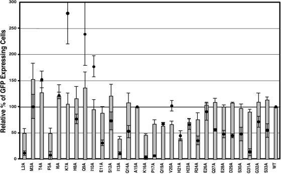
FIG. 2.
Viral entry and single-round infectivity assay. COS-1 cells were cotransfected with pTMT and pMTΔEnv expression vectors as described in Materials and Methods. At 48 h posttransfection, culture medium from cells expressing virus was filtered and normalized for reverse transcriptase activity. The normalized medium was used to infect HOS-CD4/LTR-hGFP cells. The number of GFP-expressing cells was quantitated by fluorescence-activated cell sorter analysis. The mean percentage (± the standard deviation) of GFP-expressing cells relative to wild type from three independent experiments is shown for each of the mutants (gray bars). Superimposed on the infectivity data are the fusogenicity data for each mutant in the context of a full-length Env protein expression vector (70) (filled circles).

