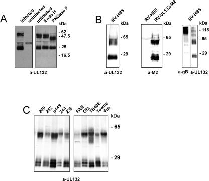
FIG. 3.
Detection of gpUL132 in HCMV infected cells and extracellular virus particles. (A) Fibroblasts were infected for 10 days with HCMV-AD169 and lysates were subjected to digestion with endoglycosidase H or PNGase F followed by immunoblot analysis using an anti-UL132 rabbit serum (a-UL132). (B) Extracellular virus particles from RV-HB5 (representing strain AD169) or a recombinant virus expressing an M2 epitope-tagged gpUL132 (RV-UL132-M2) were used for immunoblot analysis using either an anti-UL132 rabbit serum (a-UL132), an anti-M2 monoclonal antibody (a-M2), or the gB-specific monoclonal antibody 27-287 (a-gB). The analysis shown in the right panel was carried out in the absence of reducing agents. (C) Immunoblot analysis of extracellular HCMV particles purified from the tissue culture supernatant. The individual strains are indicated. The anti-UL132 rabbit serum was used for detection of gpUL132.

