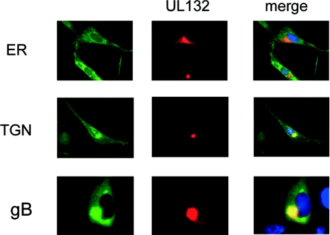
FIG. 4.
Intracellular localization of gpUL132 in infected cells. Fibroblasts were infected with the recombinant virus RV-UL132-M2 for 72 h. The intracellular localization of the individual proteins was determined by comparing the signal from Flag-specific antibody M2 (UL132) with those of antibodies or lectins specific for HCMV gB or components of the secretory pathway (endoplasmic reticulum [ER], anticalreticulin; trans-Golgi network [TGN], wheat germ agglutinin). The cellular markers and gB were developed with fluorescein isothiocyanate, and gpUL132 with Cy3-coupled anti-mouse immunoglobulin G. Yellow indicates colocalization of the signal. In the merge panel, cell nuclei are also stained blue. Magnification: panels ER and TGN, ×400; panel gB, ×630.

