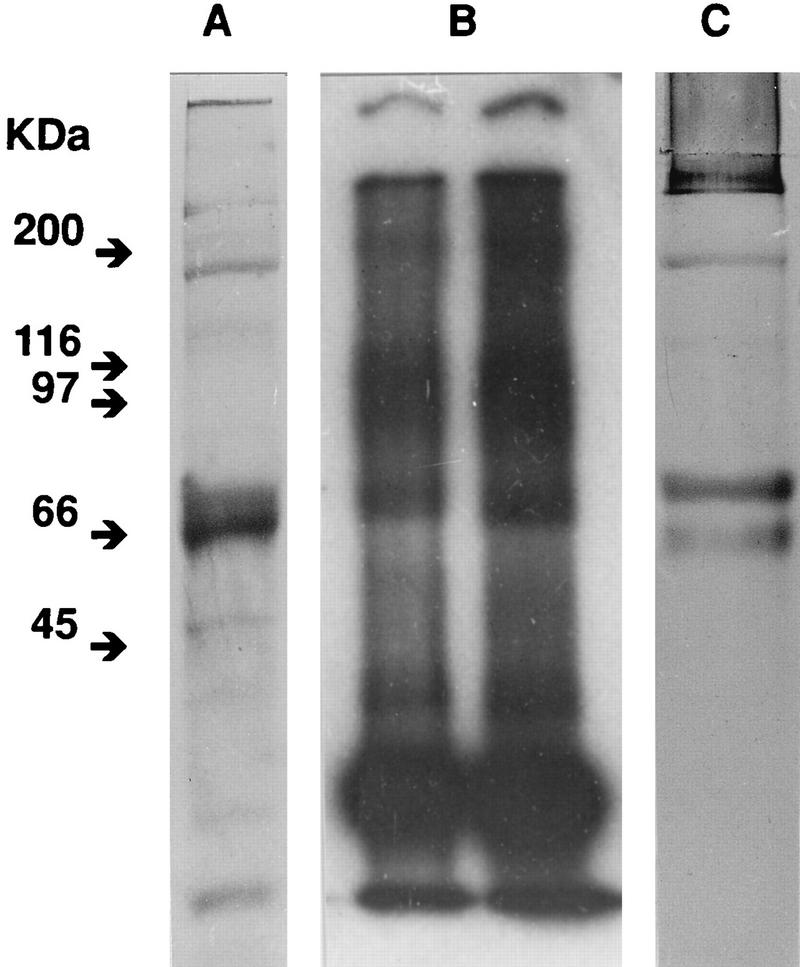
FIG. 2.
Characterization of the proteins biotinylated by reacting whole FIV with NHS-biotin. The indicated viral preparations were disrupted in SDS and subjected to SDS–10% PAGE. (A) Staining with Coomassie blue and silver of a gel obtained with biotinylated virus. (B) Autoradiography of a gel obtained with biotinylated FIV that was 125I labeled after disruption (Kodak X-Omat AR film at −70°C in the presence of Du Pont Lightning-Plus intensifying screens). (C) Staining with streptavidin of biotinylated FIV. The gel was blotted onto nitrocellulose, incubated with horseradish peroxidase-conjugated streptavidin, and examined by ECL.

