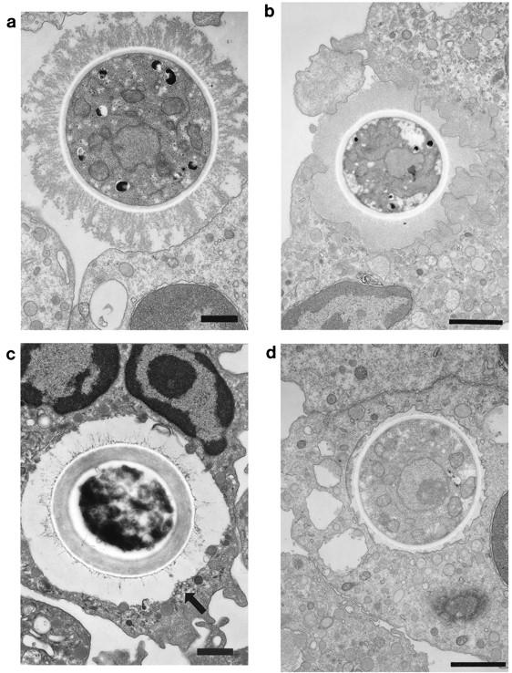
FIG. 1.

Transmission electron micrographs of sections from human PMNs incubated with 2E9- and rabbit complement-treated C. neoformans cells and processed immediately (a) or cocultured for 5 (b and c) or 30 (d) min. Bars, 1 μm. (a) Human PMN with attached C. neoformans cell. The yeast cell is seen making contact with the PMN membrane. There are no pseudopodia. (b) The yeast cell shown appears to be entering a PMN loosely surrounded by ruffled membrane from the PMN. (c) The yeast cell is seen within the cell surrounded by a large vacuolar membrane. The arrow points to a group of small vesicles beneath the vacuole. (d) A membrane-bound yeast cell is seen in a vacuole. The vacuolar membrane is tighter than that shown in panel c. FACS-separated PMNs that fluoresced had an appearance similar to that of the cells shown.
