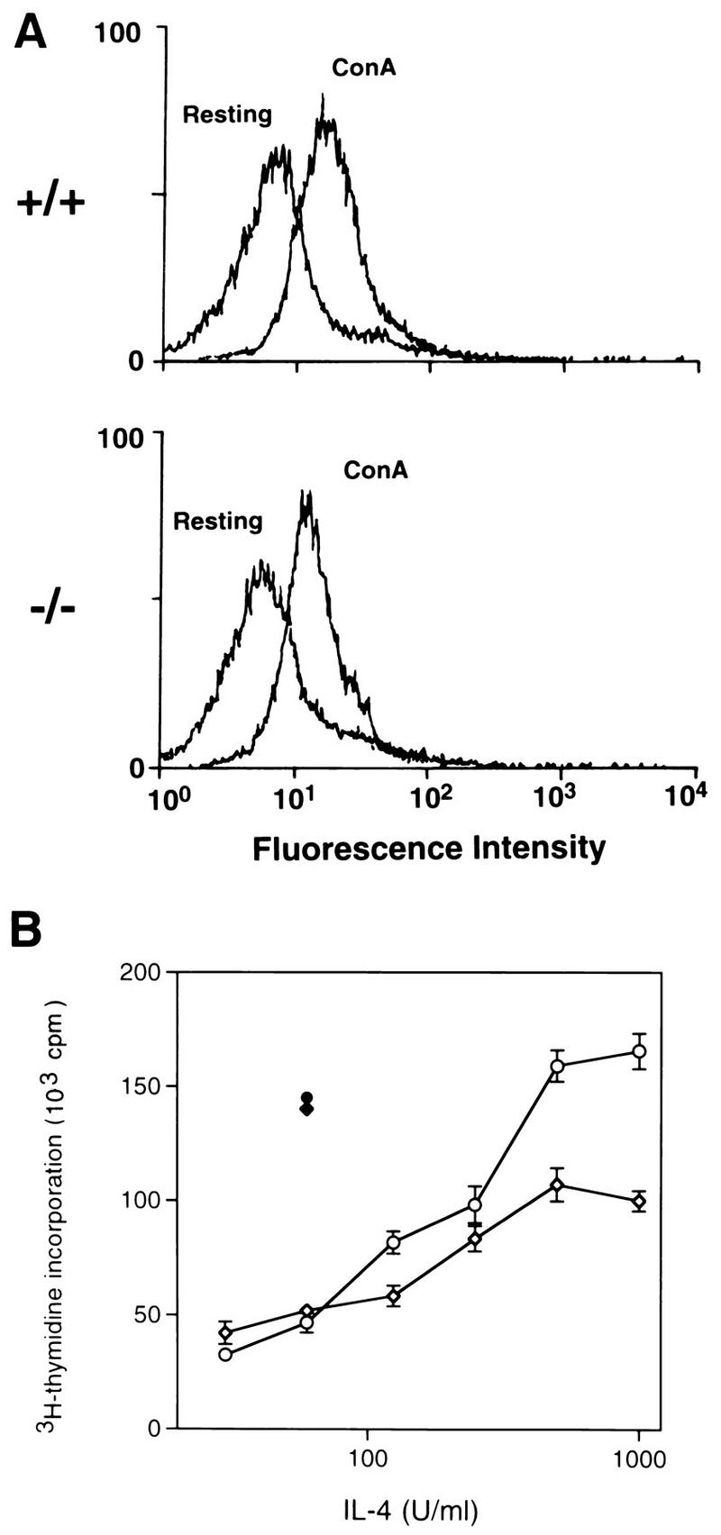
FIG. 2.

IL-4Rα expression and IL-4-induced proliferation of ConA-activated lymphocytes. (A) Flow cytometric analysis of IL-4Rα expression on control (+/+) and Stat6-deficient (−/−) lymphocytes which were either resting or activated with ConA for 48 h. (B) Control (open circles) and Stat6-deficient (open diamonds) lymphocytes activated with ConA for 48 h were assayed for proliferation in response to increasing concentrations of IL-4. The proliferation of control (solid circle) and Stat6-deficient (solid diamond) cells induced by IL-2 is also shown. Cells were pulsed with [3H]thymidine for the last 18 h of a 48-h culture period.
