
FIG. 5.
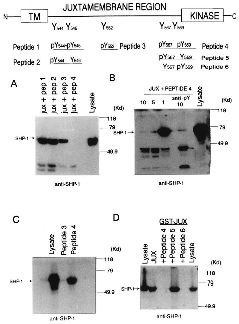
Identification of Tyr569 as the c-Kit binding site for SHP-1. (A) Phosphopeptides (12-mers) spanning the five tyrosine sites contained in the c-Kit juxtamembrane region were synthesized with tyrosines in phosphorylated or unphosphorylated states (upper diagram), and the individual phosphopeptides (10 μM) were then incubated with cell lysates (1,800 μg) from EL4 cells in the presence of 5 μg of glutathione-Sepharose–GST-JUX fusion protein. Complexes were washed four times, and the complexes and lysate protein were then subjected to SDS-PAGE followed by anti-SHP-1 immunoblotting analysis. (B) Lysates prepared from ConA-stimulated EL4 cells were incubated with glutathione-Sepharose–GST-JUX fusion proteins (5 μg) in the presence of various amounts (1, 5, or 10 μM) of phosphopeptide 4 and with 10 μM phosphopeptide 4 preincubated with anti-pTyr antibody (anti-pY). Following washing, complexes and lysate protein (500 μg) were subjected to SDS-PAGE and anti-SHP-1 immunoblotting analysis. (C) c-Kit phosphopeptides 3 and 4 were individually coupled to NHS-Sepharose beads and then incubated with ConA-treated EL4 cell lysates. Following washing, the complexes and lysate protein were resolved by SDS-PAGE and subjected to anti-SHP-1 immunoblotting analysis. (D) Glutathione-Sepharose–GST-JUX fusion protein (5 μg) was incubated with lysates from ConA-treated EL4 cells in the absence or presence of 10 μM phosphopeptide 4, 5, or 6, and the complexes and lysate proteins were subjected to SDS-PAGE and anti-SHP-1 immunoblotting analysis. In each panel, mobilities of molecular mass (MW) standards are shown on the right and the position of SHP-1 is indicated on the left.