
FIG. 7.
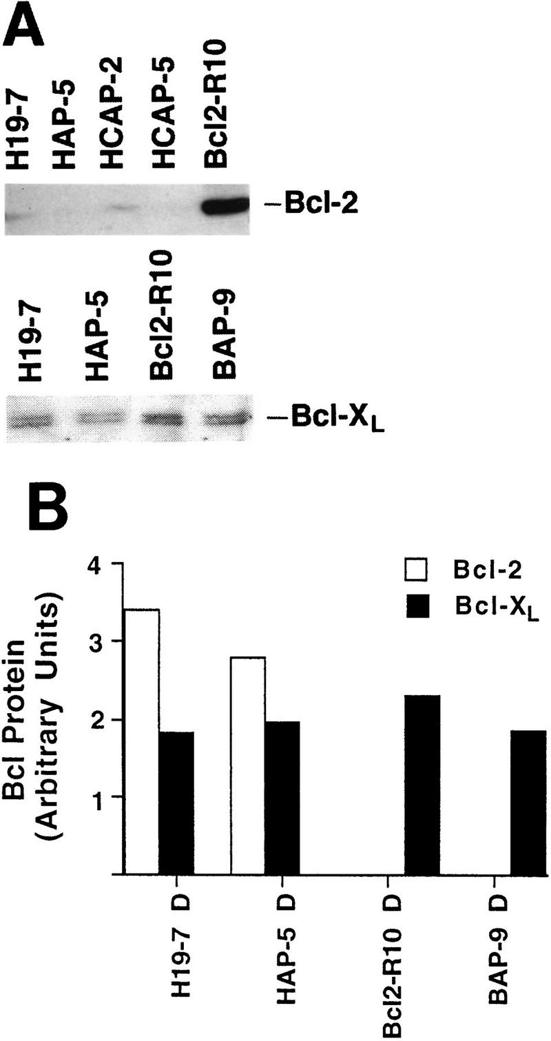
v-Akt expression does not enhance Bcl-2 or Bcl-xL protein levels in H19-7 cells. (A) Whole-cell extracts from cells differentiated for 3 days were fractionated on sodium dodecyl sulfate–10% polyacrylamide gels and immunoblotted for Bcl-2 or Bcl-xL. (B) Plot of relative Bcl-2 or Bcl-xL expression after normalization to tubulin levels. Immunoblots for Bcl-2 and Bcl-xL were reprobed with a monoclonal antibody to tubulin. Following determination of protein levels by optical density using an Ambis scanner, the amount of Bcl protein per lane was normalized to the amount of tubulin per lane. Since the Bcl-2 and Bcl-xL scans were done independently, the plot does not reflect the relative amounts of these proteins in a single cell line. D, differentiated.