FIG. 8.
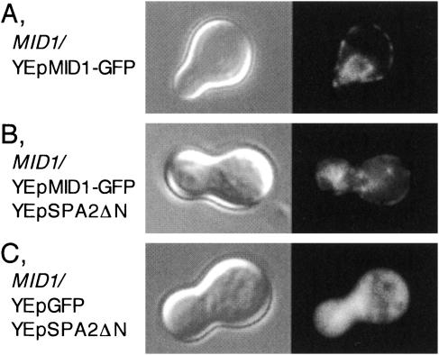
Localization of the Mid1-GFP fusion protein in Spa2ΔN-overexpressing cells. Exponentially growing MID1 cells transformed with various plasmids were incubated with 6 μM α-factor for 2 h, harvested, and observed by DIC (left panels) or fluorescence microscopy (right panels). (A) A cell transformed with YEpMID1-GFP. (B) A cell transformed with YEpMID1-GFP and YEpSPA2ΔN. (C) A cell transformed with YEpGFP and YEpSPA2ΔN. Typical examples of the cells are shown. Essentially the same results were obtained in at least two additional experiments, independently.

