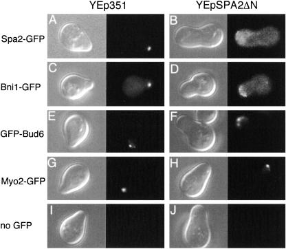
FIG. 9.
Localization of polarisome proteins in Spa2ΔN-overexpressing cells. Exponentially growing cells expressing Spa2-GFP, Bni1-GFP, GFP-Bud6, Myo2-GFP, or no GFP fusion protein were transformed with the empty vector YEp351 or YEpSPA2ΔN, incubated with 6 μM α-factor for 2 h, harvested, and subjected to microscopic analysis as described in the legend to Fig. 8. The left panel for each transformant shows DIC analysis; the right panel for each transformant shows fluorescence microscopy. (A and B) A Spa2-GFP-expressing cell (strain YKT570) with YEp351 and YEpSPA2ΔN, respectively. (C and D) A Bni1-GFP-expressing cell (YKT455) with YEp351 and YEpSPA2ΔN, respectively. (E and F) A GFP-Bud6-expressing cell (YKM14) with YEp351 and YEpSPA2ΔN, respectively. (G and H) A Myo2-GFP-expressing cell (YKT512) with YEp351 and YEpSPA2ΔN, respectively. (I and J) A cell expressing no GFP fusion protein (YKT38) with YEp351 and YEpSPA2ΔN, respectively. Typical examples of the cells are shown. Essentially the same results were obtained in at least two additional experiments, independently.

