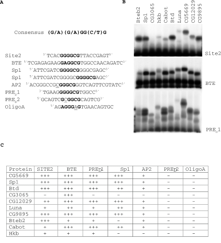
Figure 5.
DNA binding characteristics of the zinc finger regions of Drosophila SP1/KLF family members. (A) Shows the sequence of the DNA probes used in the bandshift experiments. The Sp1/KLF consensus sequence is shown. Bases that match the Sp1/KLF consensus sequence are denoted in bold type. Nucleotides that lie within the consensus but do not match it are underlined. The Sp1 oligo contains two potential Sp1/KLF binding sites. (B) Gel mobility shift assay using radioactively labeled binding site probes incubated with in vitro transcribed/translated zinc finger regions of the Drosophila Sp1/KLF family of proteins. The probe used in the bandshift is denoted beside each panel. The zinc finger regions used for the bandshift are shown at the top of the lanes. These experiments were performed simultaneously, and were repeated three times. (C) Binding of each Sp1/KLF family member to seven oligonucleotide probes. (+++) Strong binding (++) Moderate binding (+) Weak binding.

