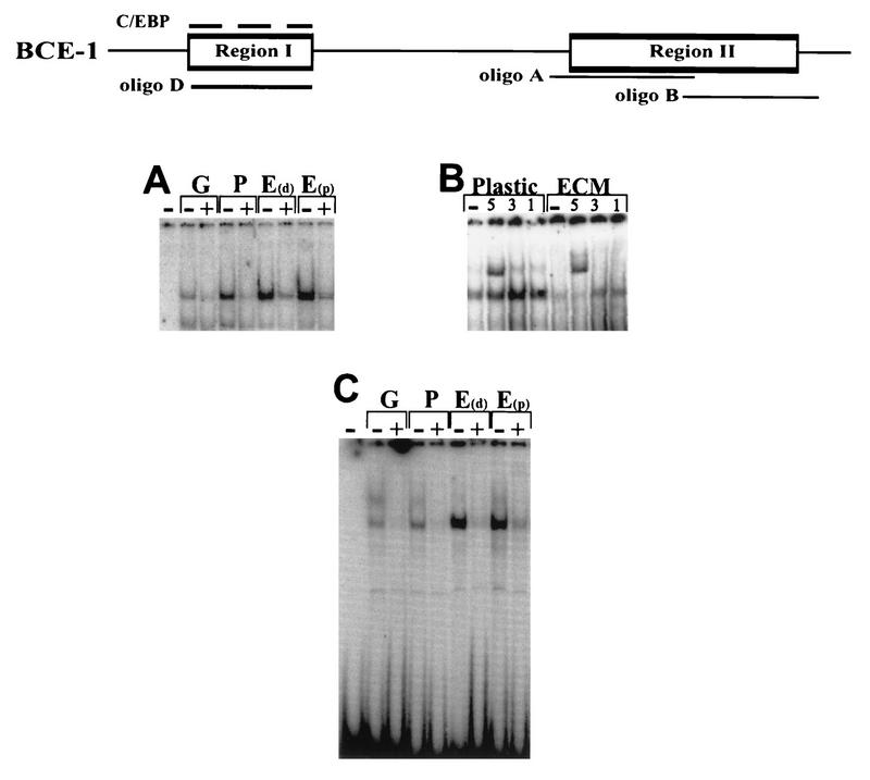
FIG. 3.
Characterization of the binding activities of proteins interacting with region II. The schematic and the extracts isolated were as described for Fig. 2. (A) EMSA using oligonucleotide A from region II as a probe with nuclear extracts from CID-9 cells as described for Fig. 2. +, lanes with a 50× molar excess of unlabeled double-stranded oligonucleotide A. (B) EMSA using oligonucleotide A with nuclear extracts from CID-9 cells cultured on plastic in IHP and ECM in IHP. −, no antibody; 5, addition of 1 μl of a 1:10 dilution of a 1:1 mixture of STAT5a and STAT5b antibodies to the binding reaction; 1 and 3, addition of 1 μl of antibody to STAT1 and STAT3, respectively. (C) EMSA using oligonucleotide B from region II as a probe. +, a 50× molar excess of unlabeled double-stranded oligonucleotide B (the quantitative differences in binding activity observed in this gel varied in at least three different EMSAs).

