FIG. 1.
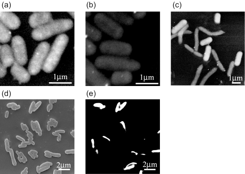
Low-vacuum (a, b, and c) and high-vacuum (d and e) SEM images of bacterial cells after ISH: E. coli hybridized with EUB338 probe (a) and NON338 probe (b), mixture of B. diminuta and E. coli hybridized with GAM42a probe (c), mixture of E. coli and A. sobria cells hybridized with ES445 probe (d and e). The same microscopic fields are shown with SE (d) and BSE images (e).

