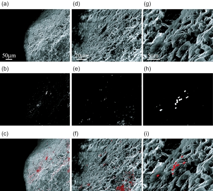
FIG. 4.
High-vacuum SEM images of bacteria attached on surface of river sediment particles detected by ISH with the EUB338 probe. The same microscopic fields are shown with SE image (a, d, and g), BSE image (b, e, and h), and their composite image (c, f, and i). The topographic information was obtained with SE images (a, d, and g), and cells hybridized with the EUB338 probe were detected with the BSE image (b, e, and h). The middle areas of the micrographs a, b, and c were magnified in micrographs d, e, and f, whose middle area was further magnified in micrographs g, h, and i. Magnifications: a, b, and c, ×250; d, e, and f, ×1,000; g, h, and i, ×5,000.

