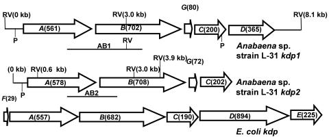
FIG. 1.
Two kdp operons from Anabaena sp. strain L-31. The arrangement of the kdp genes within the Anabaena sp. strain L-31 kdp operons is schematically depicted. The E. coli kdp operon is also shown for comparison. ORFs are shown as arrowheads indicating the direction of transcription. The expected number of amino acids for each ORF is shown in parentheses. The positions of putative promoters (P) are indicated. The chromosomal locations of the two DNA probes (AB1 and AB2) used for Southern hybridization experiments (see Fig. 2) are shown below their respective operons. The location of the EcoRV restriction enzyme site within the kdp operons is indicated (RV).

