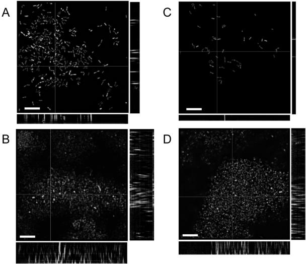
FIG. 5.
Deconvolved epifluorescence micrographs (stack average) of B. cenocepacia K56-2(pHKT2) flow cell biofilms at A) 24 h and B) 72 h and of B. cenocepacia K56-R2(pHKT2) flow cell biofilms at C) 24 h and D) 72 h. Slices were taken at 1.0-mm z-plane intervals using a Leica DMR epifluorescence microscope equipped with a focus motor at an exposure of 1.0 seconds and were volume deconvolved using Openlab 3.0.9 software. Side panels represent 1.0-μm slices in the x and y planes. Bars, 10 μm.

