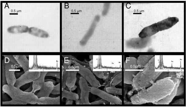
FIG. 8.
Transmission and scanning electron micrographs of S. putrefaciens under three treatments. (A and D) Cells treated with NO2−; (B and E) cells treated with Fe2+; (C and F) cells treated with both Fe2+ and NO2−. The electron-dense coating on the cells in panel C and the rough surface on the cells in panel F represent an Fe (oxy)hydroxide coating formed from the reaction between surface-bound Fe2+ and NO2−. The inset on each SEM image is an EDS point scan that corresponds to the cells in the image.

