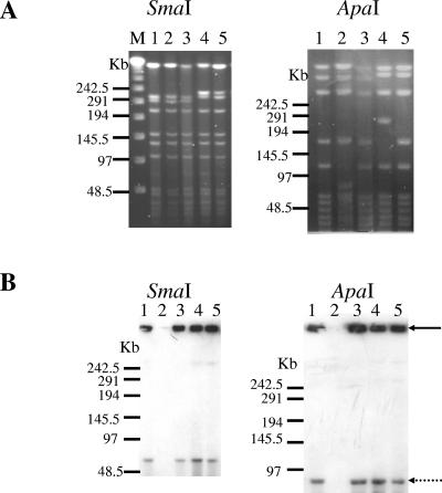
FIG. 2.
PFGE and Southern blot analysis of the six tested strains.(A) PFGE of genomic DNA after digestion with SmaI or ApaI. (B) Southern blotting of PFGE-separated fragment digested with SmaI or ApaI using a tetX-specific probe. Strains A (lane 1), G761 (lane 2), D11 (lane 3), IM1116 (lane 4), and CN1339 (lane 5) are shown. Lane M, DNA ladder PFGE marker. The black arrow indicates the position of the gel well, and the dotted arrow indicates the position of the plasmid (74 kb). Results are representative of three independent experiments.

