



Abstract
Purpose
Volar locking plate (VLP) of distal radius fractures (DRF) carries a number of surgical risks including flexor pollicis longus (FPL) rupture secondary to irritation from prominent hardware. This study reviews FPL reconstruction (with interposition graft or tendon transfer) after VLP-related rupture.
Methods
This retrospective case series reports the experience at one tertiary center with FPL reconstruction after DRF. Patients treated between January 1, 2013, and June 12, 2023, were eligible for inclusion. Preoperative radiographs were analyzed for Soong grading and fracture reduction parameters. Accessible patients (n = 7) were examined to evaluate long-term success of reconstruction.
Results
Fifteen patients (average age: 63 years) underwent VLP removal and reconstruction to restore FPL function. Rupture occurred at a median of 6 years 3 months after DRF repair (range: 1.8 months to 17 years). Eleven patients had accessible radiographs after plate fixation, all with Soong grade of 1 or 2. Eight patients underwent ring finger flexor digitorum superficialis to FPL transfer, whereas seven were treated with free tendon interposition autograft with palmaris longus (5) or flexor carpi radialis (2). One patient with systemic lupus erythematosus had a rerupture. Of the seven patients available for interview at the time of data collection, mean thumb interphalangeal joint flexion was 35°, whereas mean patient-reported outcomes measurement information system score was 48.
Conclusions
Flexor pollicis longus rupture is a known complication of VLP fixation of DRF, occurring over a wide range of time after initial fracture fixation. In cases of rupture, we have had success reconstructing FPL using tendon transfers or interposition autografts, with improvement seen after both techniques.
Type of study/level of evidence
Therapeutic IV.
Key words: Distal radius, Fracture, FPL reconstruction
Distal radius fractures are among the most common orthopedic injuries treated in the United States.1 Locking plate fixation via a volar approach is often the choice of fixation when surgical management is pursued, although this depends on multiple elements including patient factors, the fracture patterns, concomitant injuries, and surgeon preference. There are certain known risks associated with open reduction and internal fixation of distal radius fractures. These include nonspecific complications such as infection, bleeding, damage to surrounding structures, and risk of development of carpal tunnel syndrome and compartment syndrome. Unique complications exist that are specific to open treatment of distal radius fractures via a volar approach, including flexor tendon ruptures, with reports of flexor pollicis longus (FPL) rupture dating as far back as 1973 in the setting of volar buttress plate fixation.2, 3, 4, 5, 6, 7, 8, 9
Flexor pollicis longus tendon injury, although rare, is a known late complication of volar locking plate (VLP) fixation.10,11 Injuries range from cases of tenosynovitis to complete ruptures.11 The incidence of rupture has been reported to be as high as 12%, but the majority of the literature including a 2016 meta-analysis estimates that this affects less than 2.5% of patients treated with VLPs.7,8,10, 11, 12 Ruptures have been attributed not only to attrition from the distal lip of the plate primarily but also from irritation from prominent screws and fractures healing with increased dorsal tilt.3,11,13, 14, 15, 16, 17, 18, 19
After rupture from chronic attrition, primary repair of the FPL is often impossible because of tendon gap, fibrosis, synovial hypertrophy, and tendon retraction.10 Although some patients may choose to avoid surgery, many chose reconstruction including tendon grafting, tendon transfers, or thumb interphalangeal (IP) joint fusion, The authors’ preferences are to reconstruct the FPL by either using a free tendon interposition autograft using the palmaris longus (PL) or flexor carpi radialis (FCR) tendon or performing a ring finger flexor digitorum superficialis (FDS) to FPL transfer.3,20, 21, 22 This case series reports the outcomes of this treatment performed by a group of fellowship-trained hand and upper-extremity surgeons.
Materials and Methods
After obtaining institutional review board approval, we queried the surgical case records from our hand surgery service at a level 1 trauma center. We used Current Procedural Terminology codes (Appendix S1, available online on the Journal’s website at https://www.jhsgo.org) to identify patients who had undergone FPL reconstruction over the last 15 years (January 1, 2013, to June 12, 2023). After medical record review, patients were selected for study inclusion if the FPL reconstruction followed VLP fixation. Patients who had spontaneous FPL ruptures in the setting of closed reductions, diagnoses of FPL tenosynovitis without rupture, rupture of other flexor or extensor tendons, or FPL rupture of a different etiology were excluded from the study. Additionally, demographic information (ie, age, sex, diabetes, diagnosis, body mass index, ethnicity, and race), injury and surgery dates, and use of steroids was collected. Radiographic measures, including Soong grade, and fracture reduction parameters, were obtained from radiographs after fracture healing but prior to FPL reconstruction. Postoperative outcomes, specifically physical examination metrics and existing patient-reported outcomes measurement information system-upper extremity (PROMIS-UE) patient-reported outcome scores (PROs), were obtained via record review.
Eligible participants were contacted by phone, and the study was explained along with an informed consent process. Those who were reachable by phone and agreed to participate in the study met with a member of the research team (B.J.N. or A.N.W.) either in person or via secure Zoom chat. These patients completed a PROMIS short form Upper Extremity 7a version 2.0 survey.23 A test of pinch strength was performed by having the patient open a 1.5-inch standard binder clip and place over a 100-sheet pack of notepaper and then remove the clip. These materials were provided by the researchers for standardization. If the patient was able to perform to the task, they were asked to repeat it until they were unable, up to a maximum of 10 repetitions. Finally, range of motion measurements (active flexion and extension) of the affected IP joint were made using a goniometer. Interphalangeal joint flexion was deemed satisfactory if it equaled or exceeded previously reported mean functional flexion measurements (18°) for everyday tasks.24
Statistical analysis
Statistical analysis was performed to compare radiographic measurements between two reviewers (A.W. and N.G.) as well as pre- and postoperative outcomes for the FPL reconstructions. Specifically, interrater reliability was assessed using Cohen’s kappa coefficient (K) for Soong grading, and intraclass correlation coefficient (ICC) for radial inclination, ulnar variance, and dorsal tilt measurements. The difference between pre- and postoperative PROMIS-UE scores were interpreted according to a minimally clinically important difference of 3–4 points.25 The outcome measures collected during study related visits were only characterized by descriptive statistics.
Results
Record review identified 15 patients who had undergone FPL reconstruction for rupture after VLP fixation (Table 1). The patients were treated by six surgeons, and four of the 15 original VLP surgeries had been performed at our institution. Earliest rupture occurred at 7 weeks in a patient with hypothyroidism (Table 2). One patient was noted to be deceased upon chart review. Contact was successfully made with seven patients, all agreeing to participate in the follow-up study.
Table 1.
Demographics of Patients Who Underwent FPL Reconstruction After VLP Distal Radius Fixation
| Variables | N | % | Mean | SD |
|---|---|---|---|---|
| Age | - | - | 62.9 y (min 29 and max 84) | 12.9 y |
| Sex (biologic) | ||||
| Male | 9 | 60 | - | - |
| Female | 6 | 40 | - | - |
| Race | ||||
| White | 15 | 100 | - | - |
| Other | 0 | 0 | - | - |
| Ethnicity | ||||
| Hispanic | 0 | 0 | - | - |
| Non-Hispanic | 15 | 100 | - | - |
| Comorbidities | ||||
| Chronic steroid use | 0 | 0 | - | - |
| Hypothyroidism | 2 | 13 | - | - |
| Lupus | 1 | 7 | - | - |
| Timing of rupture postindex fixation | - | - | 7.4 y (min 1.75 mo and max 17 y) | 4.9 y |
| Plate Manufacturer | ||||
| Synthes | 3 | 20 | - | - |
| Acumed | 2 | 13 | - | - |
| Smith and Nephew | 1 | 7 | - | - |
| Medartis | 1 | 7 | - | - |
Table 2.
Thumb IP Joint Flexion Measurements and PROMIS-UE T-Scores at Various Time Points
| Patient | Clinic Follow-Up |
Study Follow-Up |
||||||
|---|---|---|---|---|---|---|---|---|
| Timing of Rupture From Distal Radius Fixation/mo | Pre-FPL Reconstruction PROMIS-UE T-Score | Postreconstruction PROMIS-UE T-Score at Final Clinical Follow-Up | Last Recorded IP Joint Flexion/Extension/° | Time of Last Clinic Follow-Up From Reconstruction/mo | PROMIS-UE T-Score | IP Joint Flexion/Extension/° | Time From Reconstruction/mo | |
| 1 | 58 | 28.9 | 29.1 | 18/0 | 6∗ | 29.1 | 18/0 | 6∗ |
| 2 | 74 | 32.1 | 54.9 | 48/20 | 3.5 | 49.2 | 40/0 | 22 |
| 3 | 1.75 | 21.6 | 32.1 | 35/0 | 9.5 | - | - | - |
| 4 | 144 | 55.9 | 48.3 | 40/NR | 2 | - | - | - |
| 5 | 36 | 41.9 | 43.1 | 35/NR | 5 | 41.9 | 22/−30 | 11 |
| 6 | 52 | NR | NR | 30/NR | 12 | - | - | - |
| 7 | 75 | 42.1 | 37.1 | 35/NR | 4 | - | - | - |
| 8 | 168 | 50.5 | 48.9 | 55/NR | 4 | 58.2 | 45/0 | 20 |
| 9 | 84 | 50.3 | 61 | 45/0 | 3 | - | - | - |
| 10 | 156 | 61 | 38.1 | 30/NR | 3 | - | - | - |
| 11 | 2 | NR | NR | NR/NR | 2.5 | 58.2 | 12/−30 | 126 |
| 12 | 60 | 24.4 | 33.6 | NR/NR | 16 | - | - | - |
| 13 | 96 | 42.1 | 38.1 | 50/NR | 3 | 47.7 | 60/0 | 81 |
| 14 | 204 | NR | 47.8 | 30/-5 | 5 | - | - | |
| 15 | 120 | 27.8 | 54.7 | 43/-15 | 4 | 49.3 | 50/−10 | 76 |
Abbreviation: NR, not recorded.
Represents the same follow-up appointment.
Eleven of the 15 patients had accessible wrist radiographs following plate fixation. Of these, four patients lacked calibrated imaging, precluding ulnar variance measurements, and of these four, one lacked appropriate lateral radiographs for detailed articular tilt measurements. All constructs were graded as Soong grade of 1 or 2 (Table 3), with agreement between reviewers for all except 1 construct (K = 0.79, good interrater variability, P < .05 with 100% sensitivity and 88% specificity). All patients had prominent plates that were either distal, volar, or distal, and volar to the watershed line. No patients had appreciable articular stepoff on any projection. Mean radial inclination was 16° (n = 11, ICC = 0.98), whereas ulnar variance averaged +3 mm (n = 7, ICC = 1.00) and volar tilt averaged 6° of dorsal tilt (n = 10, ICC = 1.00).
Table 3.
Soong Grading of All 11 Constructs With Accessible Prereconstruction Imaging
| Soong Grade | Number of Constructs |
|
|---|---|---|
| Reviewer 1 | Reviewer 2 | |
| 0 | 0 | 0 |
| 1 | 8 | 7 |
| 2 | 3 | 4 |
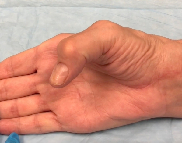
All patients were treated with VLP removal. Eight patients were treated with concomitant FDS-4 to FPL transfer; five underwent an interposed PL autograft (Fig.), whereas the remaining two underwent an interposed FCR autograft, with one being a partial harvest and the other a complete harvest. Three had an extended carpal tunnel release for exposure, and one had median nerve neurolysis after it was found to be scarred to the residual FPL tendon. There were no additional surgeries on any patient. At the time of the final documented clinic visit (median: 4 months postreconstruction), 14 patients had intact IP flexion and extension (Video S1, available online on the Journal’s website at https://www.jhsgo.org). All patients with recorded range of motion measurements achieved active thumb IP flexion of 30° or more at the last visit, except one patient who exhibited 18° of flexion (mean flexion: 38° ± 10°; Table 2). Twelve patients had documented PROMIS-UE scores from both their before surgery and the last clinic follow-up appointments. There was a small but detectable improvement between mean pre- and post-reconstruction PROMIS-UE scores (40 and 44, respectively; difference = 4). Complications included ring finger irritation (n = 1), persistent thumb IP joint stiffness (n = 1), and digital nerve neurapraxia, which subsequently resolved (n = 1). There was one case of rerupture of the tendon transfer proximal to the tenorrhaphy site at approximately 2 months after surgery, in the patient with systemic lupus erythematosus. For this patient, revision surgery was planned but never performed.
Figure.

Flexor pollicis longus reconstruction using palmaris longus interposition autograft. A Distal tenorrhaphy site. B Proximal tenorrhaphy site. C and D Thumb IP joint resting in flexion with wrist in slight extension after appropriate tensioning. Image copyright senior author C.J.D.—used with permission.
For the seven patients who participated in the follow-up study, the mean PROMIS-UE T-score at median follow-up of 22 months (range: 6–126 months) following reconstruction was 48 (Table 2). One of the seven participants was unable to place the binder clip over a 100-sheet stack of notepaper a single time. All others were able to perform the task for all 10 repetitions, demonstrating maintained good strength postreconstruction. Average active IP joint flexion and extension were 35° ± 18° and −10° ± 14°, respectively.
Discussion
In this series, median time to FPL rupture was 6.25 years after VLP fixation, with earliest rupture at 7 weeks after surgery in a patient with hypothyroidism (a previously reported risk factor for tendinopathy) and latest recorded at 17 years. For patients with accessible imaging, each construct was notably prominent (Soong grade of 1 or 2).26,27 Choice of reconstruction with tendon transfer or interposition grafting was at the discretion of the treating surgeon, with both options demonstrating largely favorable outcomes. Active thumb IP flexion of 30° or more was restored in most patients, with one outlier still achieving reported functional flexion posture of 18°.24 At a median of 22 months, majority of accessible participants continued to demonstrate functional active flexion beyond 18°. Using a gross measure of pinch function for the seven patients with long-term assessment, sufficient pinch strength was restored in six.
Active thumb IP flexion and pinch strength can be successfully restored in the setting of FPL VLP-related attritional rupture. Multiple methods exist for FPL reconstruction, ranging from end-to-end repair if acute and tendon ends remain optimal, to thumb IP joint fusion, tendon grafting,20,21,28, 29, 30 and tendon transfers. Treatment using FDS-4 to FPL transfer or a PL/FCR interposition graft has been reliably successful in our experience. In some scenarios, surgeons selected interposition grafting to optimize strength using FPL’s large, independently innervated muscle belly while avoiding donor morbidity. Others opted for FDS tendon transfer in patients requiring more precise thumb IP function for ease of achieving good tendon balance, or if the proximal FPL muscle belly was nonelastic, as in chronic ruptures. Additionally, tendon transfer requires tenorrhaphy at only one junction, which is technically less demanding and may allow quicker progress with therapy. When a free interposed tendon graft was used, PL was often the preferred choice. If absent or with improper size-matching, FCR tendon was harvested.
There are few outcome series of FPL reconstruction (Table 4). Berger et al20 reviewed four cases of PL interposition grafting after VLP-associated FPL rupture. Although they reported a significant decrease in mean IP flexion and thumb opposition when compared with contralateral control, they demonstrated favorable functional, and pain scores after reconstruction.20 The viability of PL interposition grafting as a treatment option is corroborated in two other studies. Peters et al31 performed a prospective study following 14 patients for 10 months postreconstruction. Similarly, despite a lack of return to baseline flexion and opposition, they showed comparable grip and pinch strength scores to the contralateral extremity, and excellent functional scores through the disabilities of the arm, shoulder, and hand questionnaire.31 As in our study, one patient sustained a rerupture.31 Shih et al30 also demonstrated success with return of all three patients to baseline activity. Macken et al32 built upon these results by commenting on plate prominence, with six of seven constructs attributed a Soong grade of 1, whereas one lacked available radiographs. They included differing techniques of management. Notably, they reported variable PROs but did not comment on success at the final follow-up. Our study augments the current literature by presenting combined radiographic, physical examination, and PRO data in patients who underwent interposition grafting or tendon transfer for FPL reconstruction. Through our follow-up arm, we are able to demonstrate satisfactory function and PRO scores at mid- to long-term follow-ups.
Table 4.
Comparison of Results of Case Series After FPL Reconstruction for VLP-Induced FPL Ruptures
| Case Series | Total Number of Patients | Soong Grading | Type of Reconstruction | Number of Successful Reconstructions | Number of Failed Reconstructions (ie, Reruptures) |
|---|---|---|---|---|---|
| Berger et al20 | 4 | Not reported | PL interposition autograft | 4 | 0 |
| Peters et al31 | 14 | Not reported | PL interposition autograft | 13 | 1 |
| Shih et al30 | 3 | Not reported | PL autograft | 3 | 0 |
| Macken et al32 | 7 | 1 (n = 6), unknown (n = 1) | PL interposition autograft (4), FDS-4 (1) or Brachioradialis (1) transfer, combined FDS transfer and PL autograft (1) | Not reported | Not reported |
| Current series | 15 | 1 (n = 7), 2 (n = 3), 1 or 2 (n = 1), unknown (n = 4) | PL (5) or FCR (2) interposition graft (5), FDS-4 transfer (8) | 14 | 1 |
Etiology of FPL rupture after VLP fixation has been attributed to various factors, from attrition after contact with the VLP and/or prominent screws to presence of dorsal malunion.10,33 In our patients with accessible imaging, all plates were assigned a Soong grade of 1 or 2. Thus, it is plausible that mechanical irritation from plate prominence may have contributed to FPL rupture, although it is unclear if this was because of improper positioning at time of index fixation or subsidence over time. In our practices, if the plate is considered prominent, we counsel patients to consider implant removal after union is achieved. Some surgeons have published recommendations for early removal of VLPs in patients with evidence of synovitis or irritation or in those who use steroids.3,34, 35, 36, 37 At follow-up, we find it helpful to assess for crepitus over the flexor tendons in the distal forearm during active finger motion while the wrist is in extension, as this position has been shown to maximize contact between the flexor tendons and the VLP.38 During the final follow-up, we counsel patients about possibility of delayed flexor tendon irritation and request they inform us if they develop symptoms such as swelling or weakness with pinch/grip.
This study was subject to limitations inherent to its design. Analysis of initial radiographic data was limited by incomplete or unavailable imaging for some patients. Moreover, we were unable to assess radiographs for our entire cohort of VLP constructs over the past 15 years to determine Soong grading for a control population without reported FPL rupture. As such, although suspected, we are unable to determine a causative relationship between plate positioning and rupture. Additionally, the follow-up arm of the study was limited by difficulty contacting participants, restricting inclusion to 7 of 14. The use of videochat and need for utilization of low-cost materials also limited the extent of follow-up data. However, these were practical steps taken to maximize research-specific follow-up in this rare condition with patients who had already been discharged from standard care years prior. Despite having a larger cohort than other series, our 15-patient sample size is relatively small, limiting statistical interpretation, and preventing us from comparing outcomes based on type of reconstruction. Further studies would benefit from larger numbers with long-term follow-up, particularly if intention is to compare reconstructive techniques.
Flexor pollicis longus ruptures following VLP fixation are rare but serious complications. This study supplements the limited literature, demonstrating that treatment can be successful with tendon transfer or interposition grafting. Restoring active thumb flexion is effective with either technique, and this justifies the greater time and effort cost of rehabilitation compared with IP fusion. We believe that choice should be dictated by rupture chronicity, graft availability, and size-match, discussions with patients about donor-site morbidity, and surgeon experience with tensioning either option.
Conflicts of Interest
No benefits in any form have been received or will be received related directly to this article.
Supplementary Data
Supplementary video.
References
- 1.Nellans K.W., Kowalski E., Chung K.C. The epidemiology of distal radius fractures. Hand Clin. 2012;28(2):113–125. doi: 10.1016/j.hcl.2012.02.001. [DOI] [PMC free article] [PubMed] [Google Scholar]
- 2.Margaliot Z., Haase S.C., Kotsis S.V., Kim H.M., Chung K.C. A meta-analysis of outcomes of external fixation versus plate osteosynthesis for unstable distal radius fractures. J Hand Surg Am. 2005;30(6):1185–1199. doi: 10.1016/j.jhsa.2005.08.009. [DOI] [PubMed] [Google Scholar]
- 3.Cho C.H., Lee K.J., Song K.S., Bae K.C. Delayed rupture of flexor pollicis longus after volar plating for a distal radius fracture. Clin Orthop Surg. 2012;4(4):325–328. doi: 10.4055/cios.2012.4.4.325. [DOI] [PMC free article] [PubMed] [Google Scholar]
- 4.Orbay J.L., Touhami A. Current concepts in volar fixed-angle fixation of unstable distal radius fractures. Clin Orthop Relat Res. 2006;445:58–67. doi: 10.1097/01.blo.0000205891.96575.0f. [DOI] [PubMed] [Google Scholar]
- 5.Klug R.A., Press C.M., Gonzalez M.H. Rupture of the flexor pollicis longus tendon after volar fixed-angle plating of a distal radius fracture: a case report. J Hand Surg Am. 2007;32(7):984–988. doi: 10.1016/j.jhsa.2007.05.006. [DOI] [PubMed] [Google Scholar]
- 6.Nunley J.A., Rowan P.R. Delayed rupture of the flexor pollicis longus tendon after inappropriate placement of the pi plate on the volar surface of the distal radius. J Hand Surg Am. 1999;24(6):1279–1280. doi: 10.1053/jhsu.1999.1279. [DOI] [PubMed] [Google Scholar]
- 7.Monaco N.A., Dwyer C.L., Ferikes A.J., Lubahn J.D. Hand surgeon reporting of tendon rupture following distal radius volar plating. Hand (N Y) 2016;11(3):278–286. doi: 10.1177/1558944715620792. [DOI] [PMC free article] [PubMed] [Google Scholar]
- 8.Tarallo L., Mugnai R., Zambianchi F., Adani R., Catani F. Volar plate fixation for the treatment of distal radius fractures: analysis of adverse events. J Orthop Trauma. 2013;27(12):740–745. doi: 10.1097/BOT.0b013e3182913fc5. [DOI] [PubMed] [Google Scholar]
- 9.Fuller D.J. The Ellis plate operation for Smith’s fracture. J Bone Joint Surg Br. 1973;55(1):173–178. [PubMed] [Google Scholar]
- 10.Drobetz H., Kutscha-Lissberg E. Osteosynthesis of distal radial fractures with a volar locking screw plate system. Int Orthop. 2003;27(1):1–6. doi: 10.1007/s00264-002-0393-x. [DOI] [PMC free article] [PubMed] [Google Scholar]
- 11.Cook A., Baldwin P., Fowler J.R. Incidence of flexor pollicis longus complications following volar locking plate fixation of distal radius fractures. Hand (N Y) 2020;15(5):692–697. doi: 10.1177/1558944718820956. [DOI] [PMC free article] [PubMed] [Google Scholar]
- 12.Satake H., Hanaka N., Honma R., et al. Complications of distal radius fractures treated by volar locking plate fixation. Orthopedics. 2016;39(5):e893–e896. doi: 10.3928/01477447-20160517-05. [DOI] [PubMed] [Google Scholar]
- 13.Selvan D.R., Machin D.G., Perry D., Simpson C., Thorpe P., Brown D.J. The role of fracture reduction and plate position in the aetiology of flexor pollicis longus tendon rupture after volar plate fixation of distal radius fractures. Hand (N Y) 2015;10(3):497–502. doi: 10.1007/s11552-014-9735-6. [DOI] [PMC free article] [PubMed] [Google Scholar]
- 14.Soong M., Earp B.E., Bishop G., Leung A., Blazar P. Volar locking plate implant prominence and flexor tendon rupture. J Bone Joint Surg Am. 2011;93(4):328–335. doi: 10.2106/JBJS.J.00193. [DOI] [PubMed] [Google Scholar]
- 15.Floquet A., Druart T., Lavantes P., Vendeuvre T., Delaveau A. Flexor tendon rupture after volar plating of distal radius fracture: A systematic review of the literature. Hand Surg Rehabil. 2021;40(5):535–546. doi: 10.1016/j.hansur.2021.05.008. [DOI] [PubMed] [Google Scholar]
- 16.Thorninger R., Madsen M.L., Wæver D., Borris L.C., Rölfing J.H.D. Complications of volar locking plating of distal radius fractures in 576 patients with 3.2 years follow-up. Injury. 2017;48(6):1104–1109. doi: 10.1016/j.injury.2017.03.008. [DOI] [PubMed] [Google Scholar]
- 17.Yamak K., Karahan H.G., Karatan B., Kayalı C., Altay T. Evaluation of flexor pollicis longus tendon rupture after treatment of distal radius fracture with the volar plate. J Wrist Surg. 2020;9(3):219–224. doi: 10.1055/s-0040-1702931. [DOI] [PMC free article] [PubMed] [Google Scholar]
- 18.Koo S.C., Ho S.T. Delayed rupture of flexor pollicis longus tendon after volar plating of the distal radius. Hand Surg. 2006;11(1–2):67–70. doi: 10.1142/S0218810406003048. [DOI] [PubMed] [Google Scholar]
- 19.Vasara H., Tarkiainen P., Stenroos A., et al. Higher Soong grade predicts flexor tendon issues after volar plating of distal radius fractures - a retrospective cohort study. BMC Musculoskelet Disord. 2023;24(1):271. doi: 10.1186/s12891-023-06313-0. [DOI] [PMC free article] [PubMed] [Google Scholar]
- 20.Berger P., Duerinckx J. Flexor pollicis longus tendon rupture after volar wrist plating: reconstruction with palmaris longus interposition graft. Acta Orthop Belg. 2017;83(3):467–472. [PubMed] [Google Scholar]
- 21.Arora R., Lutz M., Hennerbichler A., Krappinger D., Espen D., Gabl M. Complications following internal fixation of unstable distal radius fracture with a palmar locking-plate. J Orthop Trauma. 2007;21(5):316–322. doi: 10.1097/BOT.0b013e318059b993. [DOI] [PubMed] [Google Scholar]
- 22.Schneider L.H., Wiltshire D. Restoration of flexor pollicis longus function by flexor digitorum superficialis transfer. J Hand Surg Am. 1983;8(1):98–101. doi: 10.1016/s0363-5023(83)80065-3. [DOI] [PubMed] [Google Scholar]
- 23.Kaat A.J., Buckenmaier C.T., Cook K.F., et al. The expansion and validation of a new upper extremity item bank for the Patient-Reported Outcomes Measurement Information System® (PROMIS) J Patient Rep Outcomes. 2019;3(1):69. doi: 10.1186/s41687-019-0158-6. [DOI] [PMC free article] [PubMed] [Google Scholar]
- 24.Hume M.C., Gellman H., McKellop H., Brumfield R.H. Functional range of motion of the joints of the hand. J Hand Surg Am. 1990;15(2):240–243. doi: 10.1016/0363-5023(90)90102-w. [DOI] [PubMed] [Google Scholar]
- 25.Kazmers N.H., Qiu Y., Yoo M., Stephens A.R., Zeidan M., Zhang Y. Establishing the minimal clinically important difference for the PROMIS upper extremity computer adaptive test version 2.0 in a nonshoulder hand and upper extremity population. J Hand Surg Am. 2021;46(10):927.e1–927.e10. doi: 10.1016/j.jhsa.2021.01.023. [DOI] [PMC free article] [PubMed] [Google Scholar]
- 26.Pantazis K., Roupas N.D., Panagopoulos A., Theodoraki S., Tsintoni A., Kyriazopoulou V. Spontaneous rupture of the long head of the biceps tendon in a woman with hypothyroidism: a case report. J Med Case Rep. 2016;10(1):2. doi: 10.1186/s13256-015-0794-2. [DOI] [PMC free article] [PubMed] [Google Scholar]
- 27.Oliva F., Berardi A.C., Misiti S., Verga Falzacappa C., Iacone A., Maffulli N. Thyroid hormones enhance growth and counteract apoptosis in human tenocytes isolated from rotator cuff tendons. Cell Death Dis. 2013;4(7):e705. doi: 10.1038/cddis.2013.229. [DOI] [PMC free article] [PubMed] [Google Scholar]
- 28.Asadollahi S., Keith P.P.A. Flexor tendon injuries following plate fixation of distal radius fractures: a systematic review of the literature. J Orthop Traumatol. 2013;14(4):227–234. doi: 10.1007/s10195-013-0245-z. [DOI] [PMC free article] [PubMed] [Google Scholar]
- 29.Townsley S.H., Shin A.Y. Flexor pollicis tendon reconstruction with allograft tendon after rupture: technique and case series. Tech Hand Up Extrem Surg. 2023;27(4):225–229. doi: 10.1097/BTH.0000000000000439. [DOI] [PubMed] [Google Scholar]
- 30.Shih Y.S., Chang Y.J., Cheng Y.T., Chang C.S., Lin Y.H. Flexor pollicis longus tendon rupture: a case series on palmaris longus tendon graft reconstruction after volar plate fixation. J Surg Case Rep. 2023;2023(7) doi: 10.1093/jscr/rjad399. [DOI] [PMC free article] [PubMed] [Google Scholar]
- 31.Peters T., Caekebeke P., Duerinckx J. Reconstruction of flexor pollicis longus tendon with palmaris longus interposition graft: results in 14 patients. Acta Orthop Belg. 2022;88(4):757–760. doi: 10.52628/88.4.03. [DOI] [PubMed] [Google Scholar]
- 32.Macken A.A., Lans J., Özkan S., Kramer S., Jupiter J.B., Chen N.C. Outcomes of flexor pollicis longus reconstruction for volar plate related ruptures. J Hand Microsurg. 2021;15(3):212–218. doi: 10.1055/s-0041-1739961. [DOI] [PMC free article] [PubMed] [Google Scholar]
- 33.Monda M.K., Ellis A., Karmani S. Late rupture of flexor pollicis longus tendon 10 years after volar buttress plate fixation of a distal radius fracture: a case report. Acta Orthop Belg. 2010;76(4):549–551. [PubMed] [Google Scholar]
- 34.Cross A.W., Schmidt C.C. Flexor tendon injuries following locked volar plating of distal radius fractures. J Hand Surg Am. 2008;33(2):164–167. doi: 10.1016/j.jhsa.2007.11.011. [DOI] [PubMed] [Google Scholar]
- 35.Mathews A.L., Chung K.C. Management of complications of distal radius fractures. Hand Clin. 2015;31(2):205–215. doi: 10.1016/j.hcl.2014.12.002. [DOI] [PMC free article] [PubMed] [Google Scholar]
- 36.Tada K., Ikeda K., Shigemoto K., Suganuma S., Tsuchiya H. Prevention of flexor pollicis longus tendon rupture after volar plate fixation of distal radius fractures. Hand Surg. 2011;16(3):271–275. doi: 10.1142/S0218810411005539. [DOI] [PubMed] [Google Scholar]
- 37.Bell J.S., Wollstein R., Citron N.D. Rupture of flexor pollicis longus tendon: a complication of volar plating of the distal radius. J Bone Joint Surg Br. 1998;80(2):225–226. doi: 10.1302/0301-620x.80b2.8351. [DOI] [PubMed] [Google Scholar]
- 38.Wurtzel C.N.W., Burns G.T., Zhu A.F., Ozer K. Effects of volar tilt, wrist extension, and plate position on contact between flexor pollicis longus tendon and volar plate. J Hand Surg Am. 2017;42(12):996–1001. doi: 10.1016/j.jhsa.2017.08.008. [DOI] [PubMed] [Google Scholar]
Associated Data
This section collects any data citations, data availability statements, or supplementary materials included in this article.